door TCMVET | 9 dec. 2024 | Hondenkanker en tumoren
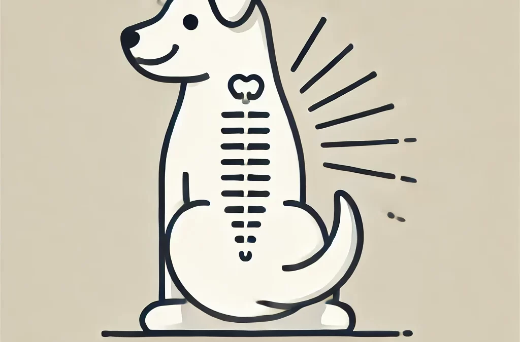
Spinale tumoren bij honden, hoewel zeldzaam, kunnen een ernstige impact hebben op hun mobiliteit en kwaliteit van leven. Deze tumoren kunnen zich ontwikkelen in of rond de wervelkolom, wat de functie van het zenuwstelsel beïnvloedt. Vroege detectie en goed beheer zijn essentieel om de beste zorg te bieden aan uw harige vriend. Hier is een uitgebreide gids over de soorten spinale tumoren bij honden en hun symptomen, oorzaken en behandelingsopties.
Veelvoorkomende soorten wervelkolomtumoren bij honden
- Intramedullaire tumoren
- Beschrijving: Deze tumoren ontstaan in het ruggenmerg zelf. Ze ontstaan vaak uit gliacellen, die het zenuwstelsel ondersteunen.
- Voorbeelden: Astrocytomen, ependymomen en oligodendrogliomen
- Symptomen: Geleidelijke zwakte, gebrek aan coördinatie en mogelijke verlamming in ernstige gevallen
- Behandeling: Chirurgie (indien mogelijk), radiotherapie en ondersteunende zorg
- Extradurale tumoren
- Beschrijving: Deze tumoren ontstaan buiten het ruggenmerg, maar in het wervelkanaal. Vaak drukken ze op het ruggenmerg en veroorzaken ze neurologische problemen.
- Voorbeelden: Osteosarcoom, fibrosarcoom en lymfoom
- Symptomen: Rugpijn, moeite met lopen en spierzwakte
- Behandeling: Afhankelijk van het type tumor: chirurgische verwijdering, chemotherapie of bestraling
- Intradurale-extramedullaire tumoren
- Beschrijving: Deze tumoren bevinden zich in het wervelkanaal, maar buiten het ruggenmerg, en groeien in de omliggende hersenvliezen of zenuwwortels.
- Voorbeelden: Meningeomen, zenuwschedetumoren (schwannomen)
- Symptomen: Pijn, gebrek aan coördinatie en mogelijke urine- of ontlastingsincontinentie
- Behandeling: Chirurgie en radiotherapie
- Wervel tumoren
- Beschrijving: Deze tumoren ontstaan in de botten van de wervelkolom en veroorzaken structurele instabiliteit en mogelijke compressie van het ruggenmerg.
- Voorbeelden: Osteosarcoom, chondrosarcoom
- Symptomen: Ernstige pijn, zwelling en moeite met staan of lopen
- Behandeling: Chirurgie, chemotherapie en pijnbestrijding
Symptoms of Spinal Tumors in Dogs
De symptomen van spinale tumoren kunnen variëren, afhankelijk van de locatie en de grootte van de tumor, maar veelvoorkomende tekenen zijn:
- Weigering om te bewegen of te spelen
- Moeilijkheden met lopen of slepen van ledematen
- Pijn of gevoeligheid in de rug of nek
- Verlies van controle over de blaas of darmen
- Plotselinge veranderingen in gedrag of houding
Oorzaken en risicofactoren
Tumoren van de wervelkolom bij honden kunnen worden veroorzaakt door:
- Genetische aanleg: Bepaalde rassen, zoals Duitse herders en golden retrievers, lopen mogelijk een hoger risico.
- Age: Oudere honden hebben meer kans op het ontwikkelen van tumoren in de wervelkolom.
- Kankermetastasen: Tumoren uit andere delen van het lichaam kunnen zich verspreiden naar de wervelkolom.
Diagnose en behandeling
Dierenartsen gebruiken verschillende methoden om spinale tumoren te diagnosticeren:
- Neurologisch onderzoek: Beoordeelt reflexen, coördinatie en pijnreactie.
- In beeld brengen: Röntgenfoto's, MRI of CT-scans om de tumor te lokaliseren en te beoordelen.
- Biopsie: Bevestigt het tumortype voor gerichte behandeling.
Behandelingsopties omvatten:
- Chirurgie: De voorkeursmethode voor toegankelijke en opereerbare tumoren.
- Bestralingstherapie: Wordt gebruikt bij inoperabele of resterende tumoren na een operatie.
- Chemotherapie: Effectief bij sommige gemetastaseerde of primaire spinale tumoren.
- Pijnbeheersing: Essentieel voor het verbeteren van de levenskwaliteit van de hond.
Uw hond ondersteunen tijdens zijn herstel
De zorg voor honden met wervelkolomtumoren omvat het volgende:
- Zorg voor een zacht, ondersteunend bed om doorligwonden te voorkomen
- Hulp bij mobiliteit door middel van harnassen of karretjes
- Het handhaven van een voedzaam dieet ter ondersteuning van de algehele gezondheid
- Regelmatige follow-up bij de dierenarts om de voortgang te bewaken
Conclusie
Spinale tumoren bij honden vereisen snelle aandacht en gespecialiseerde zorg. Hoewel de prognose afhankelijk is van het type tumor en de progressie, hebben ontwikkelingen in de diergeneeskunde behandelingen effectiever gemaakt. Door de symptomen en beschikbare opties te begrijpen, kunt u ervoor zorgen dat uw hond de beste zorg en ondersteuning krijgt.
door TCMVET | 9 dec. 2024 | Hondenkanker en tumoren
De gezondheid van de huid van honden is vaak een weerspiegeling van hun algehele welzijn, maar sommige aandoeningen kunnen zelfs voor de meest oplettende huisdiereigenaren verwarrend zijn. Een van die zeldzame aandoeningen is verhoornende epitheliomen, een type goedaardige huidtumor die vanwege zijn uiterlijk en effecten aanleiding kan geven tot bezorgdheid. Laten we dieper ingaan op deze ongewone dermatologische aandoening, de oorzaken, behandelingen en wat het tot een unieke uitdaging maakt in de gezondheidszorg voor honden.
Wat zijn verhoornende epitheliomen?
Cornificerende epitheliomen zijn goedaardige tumoren die ontstaan uit talgklieren, met name het epitheel (huidcellen) dat verantwoordelijk is voor de productie van keratine. Deze tumoren presenteren zich vaak als nodulaire, wratachtige gezwellen op de huid van een hond. Hoewel ze niet levensbedreigend zijn, mogen ze niet worden genegeerd vanwege hun potentieel om ongemak of infectie te veroorzaken.
Wat veroorzaakt verhoornende epitheliomen?
De exacte oorzaak van verhoornende epitheliomen is niet volledig bekend, maar bijdragende factoren kunnen zijn:
- Genetische aanleg: Rassen zoals Cocker Spaniels, Beagles en Siberische Husky's zijn gevoeliger voor het ontwikkelen van deze gezwellen.
- Hormonale onevenwichtigheden: De activiteit van de talgklieren kan worden beïnvloed door hormonale veranderingen, vooral bij oudere honden.
- Voedingstekorten: Slechte voeding kan leiden tot een verstoorde gezondheid van de huid, waardoor aandoeningen zoals epitheliomen kunnen verergeren.
Herkennen van de symptomen
Verhoornende epitheliomen zien er meestal als volgt uit:
- Kleine, stevige knobbeltjes met een wratachtige textuur
- Geelachtig of wasachtig van kleur door keratine-opbouw
- Gelokaliseerd rond het hoofd, de nek of de rug, maar kan overal voorkomen
- Soms gepaard gaand met roodheid of ontsteking als er een secundaire infectie optreedt
Hoewel deze gezwellen goedaardig zijn, dienen snelle veranderingen in grootte, kleur of textuur door een dierenarts te worden beoordeeld om kwaadaardige aandoeningen uit te sluiten.
Diagnose van verhoornende epitheliomen
De diagnose bestaat meestal uit:
- Fysiek onderzoek: Een dierenarts zal de grootte, locatie en het uiterlijk van de gezwellen beoordelen.
- Fijne-naaldaspiratie (FNA): Er wordt een celmonster genomen en geanalyseerd om de aard van de tumor te bevestigen.
- Biopsie: In sommige gevallen kan een biopsie nodig zijn om onderscheid te maken tussen goedaardige epitheliomen en andere huidaandoeningen of kankers.
Behandelingsopties
De behandeling hangt af van de ernst van de epitheliomen en de impact ervan op de kwaliteit van leven van uw hond.
- Toezicht houden
Bij kleine, niet-problematische gezwellen is regelmatige controle vaak voldoende.
- Zorg ervoor dat het gebied schoon en vrij van infecties blijft.
- Gebruik verzachtende plaatselijke behandelingen als uw dierenarts dit aanbeveelt.
- Chirurgisch verwijderen
Als de gezwellen ongemak, terugkerende infecties of cosmetische problemen veroorzaken, is chirurgische verwijdering een veelvoorkomende oplossing.
- Minimaal invasieve technieken zoals laserchirurgie kunnen de hersteltijd verkorten.
- Topische of systemische therapieën
- Retinoïden of vitamine A-supplementen kunnen de keratineproductie reguleren.
- Bij secundaire bacteriële infecties kunnen antibiotica worden voorgeschreven.
Innovatieve en natuurlijke benaderingen
Voor eigenaren die conventionele behandelingen willen aanvullen met holistische zorg:
- Omega-3 vetzuren: Deze kunnen ontstekingen verminderen en de algehele gezondheid van de huid bevorderen.
- Kruiden remedie: Calendula en aloë vera kunnen geïrriteerde plekken verzachten.
- Dieetaanpassingen: Een dieet rijk aan antioxidanten en hoogwaardige eiwitten ondersteunt de regeneratie van de huid.
Preventieve maatregelen
Hoewel niet alle gevallen van verhoornde epitheliomen voorkomen kunnen worden, kunnen de volgende stappen helpen om een optimale gezondheid van de huid te behouden:
- Regelmatige verzorging: Houdt de huid schoon en bevordert de vroegtijdige detectie van afwijkingen.
- Gebalanceerd dieet: Ondersteunt het immuunsysteem en vermindert de kans op huidproblemen.
- Routinematige bezoeken aan de dierenarts: Vroegtijdige interventie is essentieel voor de behandeling van elke huidaandoening.
Een unieke uitdaging in de hondendermatologie
Verhoornende epitheliomen benadrukken het belang van het begrijpen en aanpakken van zelfs zeldzame aandoeningen bij honden. Hoewel deze gezwellen goedaardig zijn, kunnen ze het comfort en uiterlijk van uw huisdier beïnvloeden, waardoor snel en effectief beheer essentieel is. Door op de hoogte te blijven en nauw samen te werken met uw dierenarts, kunt u ervoor zorgen dat uw hond gezond, gelukkig en bloeiend blijft.
door TCMVET | 8 dec. 2024 | Hondenkanker en tumoren
Opties voor het maken van afspraken
Honden likken vaak instinctief aan hun lichaam, om zichzelf te verzorgen of om ongemak te verzachten. Wanneer uw hond echter aanhoudend aan een specifiek gebied likt, zoals een tumor, kan dit duiden op een onderliggend probleem dat aandacht vereist. Dit artikel onderzoekt waarom honden tumoren likken, de mogelijke risico's en effectieve manieren om het probleem aan te pakken.
Waarom likken honden aan tumoren?
Likken is een natuurlijk gedrag voor honden, maar aanhoudend likken aan een tumor kan wijzen op verschillende onderliggende oorzaken:
- Ongemak of pijn
Tumoren, met name die ontstoken of verzweerd zijn, kunnen irritatie veroorzaken. Honden likken vaak om dit ongemak te verlichten.
- Infectie of afscheiding
Sommige tumoren kunnen bloed, pus of andere vloeistoffen produceren, die honden instinctief proberen schoon te maken door te likken.
- Nieuwsgierigheid
Honden zijn tactiele en sensorische wezens. Een groei of bult kan ongewoon aanvoelen, waardoor ze gaan onderzoeken door te likken.
- Angst of stress
Emotionele stress kan zich uiten in likgedrag. Honden kunnen zich richten op gebieden van ongemak als een zelf-kalmerend mechanisme.
Risico's van tumorlikken
Hoewel likken onschuldig lijkt, kan het tot complicaties leiden:
- Infection:Bacteriën die via likken binnenkomen, kunnen secundaire infecties veroorzaken.
- Ulceratie:Aanhoudend likken kan de huid boven de tumor beschadigen, wat verdere irritatie en bloedingen kan veroorzaken.
- Vertraagde genezing:Als de tumor behandeld of verwijderd is, kan likken het genezingsproces verstoren.
- Verspreiding van kwaadaardige cellen: In zeldzame gevallen kan irritatie door likken de verspreiding van kankercellen verergeren.
Hoe voorkom je likken?
Het is van cruciaal belang dat u onmiddellijk actie onderneemt om te voorkomen dat uw hond aan de tumor likt. Zo voorkomt u verdere complicaties.
- Raadpleeg een dierenarts
Een dierenarts moet de tumor onderzoeken om te bepalen of deze goedaardig of kwaadaardig is en welke behandeling nodig is.
- Gebruik beschermende uitrusting
Overweeg een halsband, opblaasbare halsband of bodysuit om te voorkomen dat uw hond bij de tumor komt.
- Pak de grondoorzaak aan
Afhankelijk van de diagnose kan de behandeling bestaan uit een operatie, medicatie of een combinatie van beide om het ongemak te verlichten en de tumor aan te pakken.
- Zorg voor hygiëne
Door het aangetaste gebied schoon te houden en de instructies van uw dierenarts op te volgen, kunt u infecties voorkomen en irritatie verminderen.
Ondersteun het herstel van uw hond op natuurlijke wijze
Naast conventionele behandelingen kunnen natuurlijke therapieën extra ondersteuning bieden voor de gezondheid van uw hond. Producten zoals TCMVET Baituxiao, gebaseerd op de traditionele Chinese geneeskunde, zijn speciaal ontwikkeld om de groei van tumoren te beheersen en de immuunfunctie te ondersteunen.
Huisdiereigenaren hebben positieve ervaringen gedeeld met TCMVET Baituxiao, waarbij ze verbeteringen opmerkten in de energie en het comfortniveau van hun huisdieren. Het kan een uitstekende aanvulling zijn op een uitgebreid zorgplan, maar het is essentieel om uw dierenarts te raadplegen voordat u een nieuw supplement introduceert.
Zorg voor het welzijn van uw hond
Als uw hond aan een tumor likt, is dat een duidelijk teken dat er iets mis is. Maatregelen nemen om het gedrag aan te pakken en professionele begeleiding zoeken, kan het verschil maken voor de gezondheid van uw huisdier. Het combineren van veterinaire zorg met natuurlijke supplementen zoals TCMVET Baituxiao kan de kwaliteit van leven van uw hond verbeteren en de best mogelijke ondersteuning bieden tijdens het herstel.
Door attent en proactief te blijven, kunt u ervoor zorgen dat uw hond zich op zijn gemak voelt en goed verzorgd wordt, ongeacht de uitdagingen waarmee hij te maken krijgt.
door TCMVET | 8 dec. 2024 | Hondenkanker en tumoren
Kanker is een van de meest uitdagende gezondheidsproblemen waarmee hondenbezitters te maken krijgen. De symptomen van kanker bij honden worden vaak verward met tekenen van veroudering of kleine kwalen, maar ze kunnen gemakkelijk onopgemerkt blijven totdat de ziekte is gevorderd. Dit artikel benadert de symptomen van kanker bij honden op een nieuwe manier en onderzoekt hoe natuurlijke therapieën een rol kunnen spelen bij het ondersteunen van de gezondheid van uw huisdier.
Veelvoorkomende symptomen van kanker bij honden
Kanker vroegtijdig detecteren kan een groot verschil maken in de behandelresultaten. Hier zijn enkele veelvoorkomende signalen om op te letten:
- Bulten of zwellingen:Aanhoudende bulten of ongewone zwellingen in gebieden zoals de nek, benen of buik mogen nooit genegeerd worden.
- Changes in Appetite:Een plotseling verlies van interesse in eten of moeite met eten kan duiden op onderliggende gezondheidsproblemen.
- Unexplained Weight Loss: Aanzienlijke gewichtsveranderingen zonder aanpassing van dieet of beweging kunnen wijzen op kanker.
- Gedragsveranderingen: Toenemende lusteloosheid, weinig zin om te bewegen of tekenen van ongemak kunnen wijzen op interne gezondheidsproblemen.
- Abnormale ontladingen: Aanhoudende neusuitvloeiing, bloed in de ontlasting of een vreemde geur kunnen waarschuwingssignalen zijn.
- Niet-genezende wonden:Als een wond of zweer niet binnen een redelijke tijd geneest, kan dit duiden op een dieper liggend probleem.
De rol van natuurlijke therapieën in de kankerzorg
Hoewel conventionele behandelingen zoals chirurgie, chemotherapie en bestraling vaak noodzakelijk zijn, kunnen ze duur zijn en bijwerkingen hebben. Dit is waar natuurlijke therapieën als aanvullende aanpak van pas komen. Kruiden en supplementen kunnen, wanneer zorgvuldig geselecteerd, de kwaliteit van leven van uw huisdier verbeteren en hun herstel ondersteunen.
Een dergelijk natuurlijk supplement is TCMVET Baituxiao, een formule geïnspireerd door traditionele Chinese geneeskunde. Het is ontworpen om honden te ondersteunen door tumorgroei te verminderen en de algehele balans in het lichaam te bevorderen. Huisdiereigenaren hebben verbeteringen gemeld in het energieniveau en welzijn van hun hond bij gebruik van dit product naast veterinaire zorg.
Ondersteuning van de reis van uw hond
Als uw hond een van de hierboven genoemde symptomen vertoont, raadpleeg dan onmiddellijk uw dierenarts voor een grondige diagnose. Vroege detectie, gecombineerd met een holistische benadering van de behandeling, kan het verschil maken in de reis van uw huisdier.
Het opnemen van natuurlijke therapieën zoals TCMVET Baituxiao in de verzorgingsroutine van uw hond is niet alleen een aanvulling op traditionele behandelingen, maar biedt ook extra ondersteuning voor hun algehele gezondheid. Door geïnformeerd en proactief te blijven, kunt u ervoor zorgen dat uw harige metgezel de best mogelijke zorg krijgt.

door TCMVET | 7 dec. 2024 | Hondenkanker en tumoren
Transitioneel celcarcinoom (TCC) is het meest voorkomende type urineblaaskanker bij honden en vormt vaak een grote uitdaging voor zowel huisdieren als hun eigenaren. Ondanks de agressieve aard ervan kunnen vroege detectie en een proactieve benadering van de behandeling de kwaliteit van leven van een hond helpen verbeteren.
Wat is overgangscelcarcinoom?
TCC is een kwaadaardige kanker die doorgaans ontstaat in de epitheliale bekleding van de blaas, met name in het trigonale gebied, waar de urethra en ureters samenkomen. Hoewel het voornamelijk de blaas aantast, kan het zich verspreiden naar de urethra, prostaat, lymfeklieren en andere organen als het onbehandeld blijft.
Welke honden lopen risico?
Hoewel TCC elke hond kan treffen, zijn bepaalde rassen er vatbaar voor, waaronder:
- Schotse Terriërs (hoogste risico)
- Shetland-herdershonden
- West Highland White Terriërs
- Beagles
Vrouwelijke honden en oudere honden hebben een grotere kans op TCC, hoewel de aandoening niet uitsluitend bij deze groepen voorkomt.
Symptomen waar u op moet letten
Vroege tekenen van TCC kunnen lijken op veelvoorkomende urineweginfecties, wat de diagnose lastig maakt. Belangrijke symptomen zijn:
- Moeite met plassen (dysurie)
- Verhoogde frequentie van urineren (pollakisurie)
- Bloed in de urine (hematurie)
- Urine-incontinentie
- Moeilijkheden met plassen of volledige blokkade
Naarmate de ziekte vordert, kunnen symptomen als lusteloosheid, gewichtsverlies en buikpijn optreden.
Hoe wordt TCC vastgesteld?
Om TCC te diagnosticeren, is een combinatie van tests nodig om andere aandoeningen uit te sluiten en de aanwezigheid van kanker te bevestigen:
- Urineonderzoek: Helpt bloed, bacteriën of abnormale cellen te detecteren.
- In beeld brengen: Met behulp van een echo of röntgenfoto kunnen blaastumoren zichtbaar worden gemaakt.
- Cystoscopy: Directe visualisatie van de blaas voor het nemen van een biopsie.
- BRAF-mutatietest: Een niet-invasieve urinetest die mutaties detecteert die verband houden met TCC.
Een vroege en nauwkeurige diagnose is essentieel voor de implementatie van een effectief behandelplan.
Behandelingsopties
Hoewel TCC zelden te genezen is, zijn er verschillende behandelingen die de ziekte onder controle kunnen houden en de kwaliteit van leven van de hond kunnen verbeteren:
- Medicijnen:
- Niet-steroïde ontstekingsremmers (NSAID's) zoals piroxicam worden vaak voorgeschreven vanwege hun antikankereigenschappen.
- Chemotherapeutische middelen zoals mitoxantron of vinblastine kunnen alleen of in combinatie met NSAID's worden gebruikt.
- Chirurgie:
- Als de tumor lokaal is, is chirurgische verwijdering een optie. De locatie van de tumor in het trigonale gebied maakt dit echter vaak lastig.
- Bestralingstherapie:
- Kan helpen tumoren te verkleinen en symptomen te verlichten wanneer een operatie niet mogelijk is.
- Palliatieve zorg:
- Gericht op het behoud van comfort door middel van pijnbestrijding en het aanpakken van urinewegobstructies.
Leven met TCC: wat eigenaren moeten weten
Het managen van een hond met TCC omvat regelmatige veterinaire controles en het monitoren van symptomen. Hier zijn enkele tips voor huisdiereigenaren:
- Dieet en hydratatie: Zorg voor een uitgebalanceerd dieet en zorg ervoor dat de hond voldoende vocht binnenkrijgt om de gezondheid van de urinewegen te ondersteunen.
- Medicijnen: Dien de voorgeschreven medicijnen consequent toe en meld eventuele bijwerkingen aan uw dierenarts.
- Observatie: Let op veranderingen in het urineerpatroon of tekenen van ongemak.
- Emotionele steun: TCC kan stressvol zijn voor zowel het huisdier als de eigenaar. Zoek daarom steun bij uw dierenarts of bij dierenverenigingen.
Prognose
De prognose voor honden met TCC varieert afhankelijk van het stadium van de ziekte en het behandelplan. Met de juiste zorg kunnen veel honden maanden of zelfs meer dan een jaar na de diagnose comfortabel leven. Vroege detectie en een proactieve aanpak kunnen een groot verschil maken in het verlengen van het leven van een hond en het behouden van hun kwaliteit van leven.
Laatste gedachten
Hoewel transitioneel celcarcinoom een ernstige diagnose is, bieden ontwikkelingen in de diergeneeskunde hoop en opties voor huisdiereigenaren. Door de ziekte te begrijpen en nauw samen te werken met uw dierenarts, kunt u ervoor zorgen dat uw hond de best mogelijke zorg krijgt.
door TCMVET | 7 dec. 2024 | Hondenkanker en tumoren
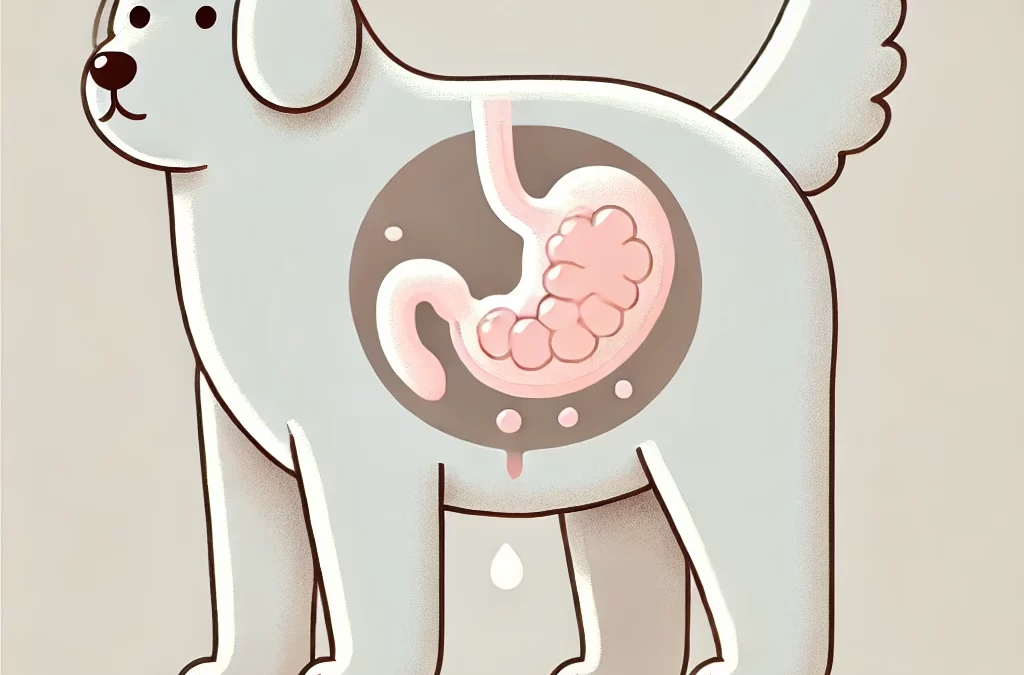
Het ontdekken van een grote abdominale massa bij een hond kan alarmerend zijn voor elke huisdiereigenaar. Hoewel het natuurlijk is om je zorgen te maken, kan het begrijpen van de mogelijke oorzaken, symptomen en behandelingsopties je in staat stellen om weloverwogen beslissingen te nemen over de gezondheid van je hond.
Wat is een abdominale massa?
Een abdominale massa is een abnormale groei in het maaggebied die kan ontstaan uit organen zoals de lever, milt, darmen of zelfs omliggende weefsels. Deze massa's kunnen aanzienlijk variëren in grootte en kunnen goedaardig (niet-kankerachtig) of kwaadaardig (kankerachtig) zijn.
Veelvoorkomende oorzaken van abdominale massa's
- Goedaardige gezwellen
- Lipomen: Vetgezwellen die over het algemeen ongevaarlijk zijn.
- Cysten: Met vocht gevulde zakjes die in de loop van de tijd kunnen groeien, maar doorgaans niet gevaarlijk zijn.
- Kwaadaardige tumoren
- Hemangiosarcoom: Een veelvoorkomende vorm van kanker die de milt aantast.
- Lymfoom: tast de lymfeklieren aan en kan zich verspreiden naar de buik.
- Carcinomen: Tumoren die ontstaan uit epitheelweefsels van inwendige organen.
- Andere oorzaken
- Abcessen: Infecties die leiden tot met pus gevulde holtes.
- Vergroting van organen: Aandoeningen zoals leverziekten kunnen lijken op massagroei.
Symptomen waar u op moet letten
Vroege detectie kan een significante impact hebben op de uitkomsten. Let op:
- Gezwollen of opgezwollen buik
- Gewichtsverlies of verminderde eetlust
- Braken of diarree
- Lethargie of tekenen van ongemak
- Moeilijk ademhalen door druk in de buik
Diagnose
Het diagnosticeren van een abdominale massa vereist de expertise van een dierenarts. Procedures kunnen het volgende omvatten:
- Fysiek onderzoek: Palperen van de buik om ongewone gezwellen te detecteren
- Beeldvormingstests: Echografie of röntgenfoto's om de grootte, locatie en aard van de massa te bepalen
- Biopsie of fijne naaldaspiratie: Het verzamelen van weefselmonsters voor analyse
- Blood Tests: Controleren op gerelateerde gezondheidsproblemen
Behandelingsopties
De behandeling is afhankelijk van het type en de locatie van de massa:
- Chirurgisch verwijderen: Vaak de eerste actielijn voor operationele massa's
- Chemotherapie of bestraling: Wordt gebruikt bij kwaadaardige gezwellen, vooral als een operatie niet mogelijk is
- Palliatieve zorg: Richt zich op het behoud van comfort als de aandoening ongeneeslijk is
Proactieve stappen voor huisdiereigenaren
- RoutinecontrolesRegelmatige bezoeken aan de dierenarts kunnen helpen om problemen vroegtijdig op te sporen
- Houd de symptomen in de gaten: Houd veranderingen in het gedrag of de eetlust van uw hond in de gaten
- Goede voeding: Een evenwichtig dieet ondersteunt de algehele gezondheid en het herstel
- Tweede meningen: Aarzel niet om bij complexe gevallen een andere dierenarts te raadplegen
Grote buikmassa's bij honden zijn een serieuze zorg, maar met tijdige veterinaire zorg en goed management kunnen veel honden goede resultaten behalen. Werk altijd nauw samen met uw dierenarts om de beste handelwijze voor uw harige vriend te bepalen.
