執筆者 TCMVET | 2024年12月9日 | 犬の癌と腫瘍
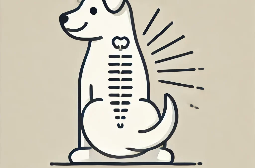
犬の脊髄腫瘍は稀ではありますが、犬の運動能力や生活の質に重大な影響を及ぼす可能性があります。これらの腫瘍は脊柱の内部または周囲に発生し、神経系の機能に影響を及ぼす可能性があります。愛犬に最善のケアを提供するには、早期発見と適切な管理が不可欠です。ここでは、犬の脊髄腫瘍の種類とその症状、原因、治療オプションに関する包括的なガイドを紹介します。
犬の脊椎腫瘍の一般的な種類
- 髄内腫瘍
- 説明: これらの腫瘍は脊髄自体に発生し、神経系を支えるグリア細胞から発生することが多いです。
- 例: 星細胞腫、上衣腫、乏突起膠腫
- 症状: 徐々に衰弱し、協調運動障害が起こり、重症の場合は麻痺が起こる可能性もある
- 治療だ: 手術(可能な場合)、放射線療法、支持療法
- 硬膜外腫瘍
- 説明: これらの腫瘍は脊髄の外側、脊柱管内に発生し、脊髄を圧迫して神経系の問題を引き起こすことがよくあります。
- 例: 骨肉腫、線維肉腫、リンパ腫
- 症状: 腰痛、歩行困難、筋力低下
- 治療だ: 腫瘍の種類に応じて、外科的切除、化学療法、または放射線療法
- 硬膜内髄外腫瘍
- 説明: これらの腫瘍は脊柱管内、脊髄の外側に位置し、周囲の髄膜または神経根で増殖します。
- 例: 髄膜腫、神経鞘腫瘍(シュワン細胞腫)
- 症状: 痛み、協調運動障害、尿失禁や便失禁の可能性
- 治療だ: 手術と放射線治療
- 脊椎腫瘍
- 説明: これらの腫瘍は脊椎の骨から発生し、構造的不安定性を引き起こし、脊髄を圧迫する可能性があります。
- 例: 骨肉腫、軟骨肉腫
- 症状: 激しい痛み、腫れ、立ち上がったり歩いたりするのが困難
- 治療だ: 手術、化学療法、疼痛管理
Symptoms of Spinal Tumors in Dogs
脊椎腫瘍の症状は腫瘍の位置と大きさによって異なりますが、一般的な兆候は次のとおりです。
- 動くことや遊ぶことを嫌がる
- 歩行困難または手足を引きずる
- 背中や首の痛みや敏感さ
- 排尿または排便のコントロールの喪失
- 行動や姿勢の突然の変化
原因と危険因子
犬の脊椎腫瘍は、以下の原因で発生することがあります。
- 遺伝的素因: ジャーマンシェパードやゴールデンレトリバーなどの特定の犬種は、リスクが高い可能性があります。
- Age: 高齢犬は脊椎腫瘍を発症する可能性が高くなります。
- がんの転移: 体の他の部分の腫瘍が脊椎に転移する可能性があります。
診断と治療
獣医師は脊椎腫瘍を診断するためにいくつかの方法を使用します。
- 神経学的検査: 反射、協調性、痛みの反応を評価します。
- イメージング: 腫瘍の位置を特定し評価するためのX線、MRI、またはCTスキャン。
- 生検: 標的治療のための腫瘍の種類を確認します。
治療の選択肢には以下のものがあります:
- 手術だ: アクセス可能で手術可能な腫瘍に推奨される方法です。
- 放射線療法: 手術不能または手術後の残存腫瘍に使用されます。
- 化学療法: 一部の転移性または原発性脊椎腫瘍に有効です。
- 疼痛管理: 犬の生活の質を向上させるために不可欠です。
犬の回復をサポートする
脊髄腫瘍のある犬のケアには以下が含まれます。
- 褥瘡を防ぐために柔らかくてサポート力のあるベッドを提供する
- ハーネスやカートによる移動の補助
- 全体的な健康をサポートするために栄養価の高い食事を維持する
- 定期的に獣医師によるフォローアップを行い、進捗状況を監視する
結論
犬の脊髄腫瘍には、迅速な対応と専門的なケアが必要です。予後は腫瘍の種類と進行度によって異なりますが、獣医学の進歩により、治療はより効果的になっています。症状と利用可能な選択肢を理解することで、愛犬が最善のケアとサポートを受けられるようになります。
執筆者 TCMVET | 2024年12月9日 | 犬の癌と腫瘍
犬の皮膚の健康は、その全体的な健康状態を反映することが多いのですが、最も注意深い飼い主でさえも困惑してしまうような症状もあります。そのような稀な症状の1つは 角化上皮腫は、その外観と影響から懸念される良性の皮膚腫瘍の一種です。この珍しい皮膚疾患、その原因、治療法、そして犬のヘルスケアにおけるこの疾患のユニークな課題について詳しく見ていきましょう。
角化上皮腫とは何ですか?
角化上皮腫は、皮脂腺、特にケラチン生成を担う上皮(皮膚細胞)から発生する良性腫瘍です。この腫瘍は、犬の皮膚に結節状のイボのような腫瘍として現れることがよくあります。命にかかわるものではありません。しかし、不快感や感染症を引き起こす可能性があるため、無視すべきではありません。
角化上皮腫の原因は何ですか?
角化上皮腫の正確な原因は完全には解明されていませんが、次のような要因が考えられます。
- 遺伝的素因: コッカースパニエル、ビーグル、シベリアンハスキーなどの犬種は、こうした腫瘍を発症する傾向があります。
- ホルモンの不均衡: 皮脂腺の活動は、特に高齢の犬ではホルモンの変化によって影響を受けることがあります。
- 食事不足: 栄養不足は皮膚の健康状態の不均衡につながり、上皮腫などの症状を悪化させる可能性があります。
症状を認識する
角化上皮腫は典型的には以下のように現れます。
- イボのような質感を持つ小さくて硬い結節
- ケラチンの蓄積により黄色またはワックス状になる
- 頭、首、背中の周囲に限局するが、どこにでも発生する可能性がある
- 二次感染が起こると、赤みや炎症を伴うことがあります。
これらの腫瘍は良性ですが、大きさ、色、または質感の急激な変化については、悪性腫瘍を除外するために獣医師による評価が必要です。
角化上皮腫の診断
診断には通常、以下が含まれます。
- 身体検査: 獣医師は腫瘍の大きさ、位置、外観を評価します。
- 穿刺吸引法(FNA): 細胞のサンプルを抽出して分析し、腫瘍の性質を確認します。
- 生検: 場合によっては、良性上皮腫と他の皮膚疾患または癌を区別するために生検が必要になることがあります。
治療の選択肢
治療法は、上皮腫の重症度と犬の生活の質に与える影響に応じて異なります。
- モニタリング
小さくて問題のない腫瘍の場合は、定期的なモニタリングで十分な場合がよくあります。
- 患部が清潔で感染がないことを確認してください。
- 獣医師から勧められた場合は、鎮静効果のある局所治療薬を使用してください。
- 外科的除去
腫瘍が不快感、再発性感染症、または美容上の懸念を引き起こしている場合は、外科的切除が一般的な解決策です。
- レーザー手術のような低侵襲技術により、回復時間を短縮できます。
- 局所療法または全身療法
- レチノイドやビタミン A サプリメントはケラチンの生成を調節することができます。
- 二次的な細菌感染に対しては抗生物質が処方されることがあります。
革新的かつ自然なアプローチ
従来の治療をホリスティックケアで補いたいと考えている飼い主様へ:
- オメガ3脂肪酸: これらは炎症を軽減し、全体的な肌の健康を促進します。
- 薬草: カレンデュラとアロエベラは炎症を起こした部分を和らげます。
- 食事の調整: 抗酸化物質と高品質のタンパク質を豊富に含む食事は、肌の再生をサポートします。
予防措置
角質化上皮腫のすべての症例を予防できるわけではありませんが、以下の手順で最適な皮膚の健康を維持することができます。
- 定期的なグルーミング: 肌を清潔に保ち、異常の早期発見を促します。
- バランスの取れた食事: 免疫システムをサポートし、皮膚の問題の可能性を減らします。
- 定期的な獣医の診察: 早期介入があらゆる皮膚疾患の管理の鍵となります。
犬の皮膚科におけるユニークな課題
角化上皮腫は、犬のまれな病気であっても理解し対処することの重要性を浮き彫りにします。これらの腫瘍は良性ですが、ペットの快適さや外見に影響を与える可能性があるため、迅速かつ効果的な管理が不可欠です。情報を入手し、獣医師と緊密に連携することで、愛犬が健康で幸せで元気に過ごせるようにすることができます。

執筆者 TCMVET | 2024年12月8日 | 犬の癌と腫瘍
tmentオプション
犬は、身だしなみを整えるため、または不快感を和らげるため、本能的に体を舐めることがよくあります。しかし、犬が腫瘍などの特定の場所を執拗に舐める場合は、注意が必要な根本的な問題を示している可能性があります。この記事では、犬が腫瘍を舐める理由、潜在的なリスク、およびこの問題に対処する効果的な方法について説明します。
犬はなぜ腫瘍を舐めるのでしょうか?
犬にとって舐めることは自然な行動ですが、腫瘍を執拗に舐める場合は、いくつかの根本的な理由が考えられます。
- 不快感または痛み
腫瘍、特に炎症を起こしたり潰瘍ができたりしたものは、刺激を引き起こす可能性があります。犬は、この不快感を和らげるために舐めることがよくあります。
- 感染または分泌物
腫瘍によっては、血液、膿、その他の体液が出ることがありますが、犬は本能的に舐めてそれをきれいにしようとします。
- 好奇心
犬は触覚と感覚に敏感な生き物です。腫瘍やしこりがあると、犬は異常に感じて舐めて調べようとすることがあります。
- 不安やストレス
感情的なストレスは舐める行動として現れることがあります。犬は、自己鎮静メカニズムとして、不快な部分に集中することがあります。
腫瘍を舐めるリスク
舐めることは無害に思えるかもしれませんが、合併症を引き起こす可能性があります。
- Infection舐めることで細菌が入り込み、二次感染を引き起こす可能性があります。
- 潰瘍: 舐め続けると腫瘍の上の皮膚が傷つき、さらなる炎症や出血を引き起こす可能性があります。
- 治癒の遅れ: 腫瘍が治療または除去された場合、舐めると治癒プロセスが妨げられる可能性があります。
- 悪性細胞の拡散: まれに、舐めることによる刺激が癌細胞の拡散を悪化させる場合があります。
舐めるのを防ぐ方法
さらなる合併症を避けるためには、犬が腫瘍を舐めないようにすぐに行動を起こすことが重要です。
- 獣医に相談する
獣医師は腫瘍を検査して良性か悪性かを判断し、適切な治療方針を勧める必要があります。
- 保護具を使用する
犬が腫瘍に近づかないように、エリザベスカラー、膨張式カラー、またはボディスーツの使用を検討してください。
- 根本原因に対処する
診断に応じて、不快感を軽減し腫瘍に対処するために、手術、投薬、またはその両方の組み合わせによる治療が行われる場合があります。
- 衛生を保つ
患部を清潔に保ち、獣医の指示に従うことで、感染を防ぎ、炎症を軽減することができます。
愛犬の回復を自然にサポート
従来の治療法に加えて、自然療法は犬の健康をさらにサポートすることができます。 TCMVET 白頭霄伝統的な中国医学に基づいて、腫瘍の成長を抑制し、免疫機能をサポートするように特別に処方されています。
ペットの飼い主は、TCMVET Baituxiao を使用したことでペットのエネルギーと快適さが向上したと肯定的な感想を述べています。これは総合的なケア プランに追加するのに最適ですが、新しいサプリメントを導入する前には必ず獣医師に相談してください。
愛犬の健康を守る
愛犬が腫瘍を舐めている場合は、何かがおかしいという明らかな兆候です。その行動に対処するための措置を講じ、専門家の指導を求めることで、ペットの健康に大きな違いが生まれます。獣医によるケアと TCMVET Baituxiao などの天然サプリメントを組み合わせることで、愛犬の生活の質が向上し、回復中に最大限のサポートを提供できます。
注意深く積極的に行動することで、犬がどんな困難に直面しても、犬が快適に過ごし、世話をされている状態を保つことができます。
執筆者 TCMVET | 2024年12月8日 | 犬の癌と腫瘍
がんは、犬の飼い主が直面する最も困難な健康問題の一つです。犬のがんの症状は、老化の兆候や軽い病気と間違われることが多く、病気が進行するまで気付かれないことがよくあります。この記事では、犬のがんの症状を理解するための新しいアプローチを採用し、自然療法がペットの健康をサポートする上でどのような役割を果たすことができるかを探ります。
犬の癌の一般的な症状
がんを早期に発見すると、治療結果に大きな違いが生まれます。注意すべき一般的な兆候は次のとおりです。
- しこりや腫れ首、脚、腹部などの部位に持続的なしこりや異常な腫れがある場合は、決して無視しないでください。
- Changes in Appetite突然食べ物への興味がなくなったり、食べるのが困難になったりする場合は、根本的な健康上の問題が示唆されることがあります。
- Unexplained Weight Loss食事や活動を調整せずに体重が大幅に変化した場合は、がんの兆候である可能性があります。
- 行動の変化無気力の増加、運動への嫌悪、または不快感の兆候は、内部の健康問題を示している可能性があります。
- 異常放電: 持続的な鼻水、便に血が混じっている、または異常な臭いがする場合は警告サインである可能性があります。
- 治癒しない傷: 傷や痛みが適切な時間内に治らない場合は、より深刻な問題がある可能性があります。
がん治療における自然療法の役割
手術、化学療法、放射線療法などの従来の治療は多くの場合必要ですが、費用がかかり、副作用を伴うこともあります。そこで、補完的なアプローチとして自然療法が役立ちます。ハーブやサプリメントを慎重に選択すれば、ペットの生活の質を高め、回復をサポートすることができます。
そのような天然サプリメントの一つは TCMVET 白頭霄は、伝統的な中国医学にヒントを得た処方です。腫瘍の成長を抑え、体全体のバランスを促進することで犬をサポートするように設計されています。ペットの飼い主は、獣医によるケアと併せてこの製品を使用すると、犬のエネルギーレベルと健康状態が改善したと報告しています。
あなたの愛犬の旅をサポートする
愛犬が上記の症状のいずれかを示している場合は、すぐに獣医に相談して、徹底的な診断を受けてください。早期発見と総合的な治療アプローチを組み合わせることで、ペットの人生に大きな違いが生まれます。
TCMVET Baituxiao のような自然療法を犬のケア ルーチンに取り入れることは、従来の治療法を補完するだけでなく、犬の全体的な健康をさらにサポートすることにもなります。情報を入手し、積極的に行動することで、愛犬が可能な限り最高のケアを受けられるようにすることができます。

執筆者 TCMVET | 2024年12月7日 | 犬の癌と腫瘍
移行上皮癌 (TCC) は、犬の膀胱癌の中で最も一般的なタイプであり、ペットとその飼い主の両方にとって大きな問題となることがよくあります。その悪性度にもかかわらず、早期発見と積極的な治療アプローチは、犬の生活の質を向上させるのに役立ちます。
移行上皮癌とは何ですか?
TCC は、通常、膀胱の上皮層、特に尿道と尿管が交わる膀胱三角部に発生する悪性癌です。主に膀胱に影響を及ぼしますが、治療せずに放置すると、尿道、前立腺、リンパ節、その他の臓器に転移する可能性があります。
どの犬が危険にさらされているのでしょうか?
TCC はどの犬にも発生する可能性がありますが、次のような特定の犬種は発症しやすい傾向があります。
- スコティッシュ・テリア(最もリスクが高い)
- シェットランド・シープドッグ
- ウエストハイランドホワイトテリア
- ビーグル
TCC はメスの犬や高齢の犬に発生する可能性が高くなりますが、この症状はこれらのグループに限ったものではありません。
注意すべき症状
TCC の初期症状は一般的な尿路感染症に似ているため、診断が困難です。主な症状は次のとおりです。
- 排尿困難(排尿困難)
- 排尿回数の増加(頻尿)
- 尿に血が混じる(血尿)
- 尿失禁
- 排尿困難または排尿障害
病気が進行すると、無気力、体重減少、腹痛などの症状が現れることがあります。
TCC はどのように診断されますか?
TCC の診断には、他の病状を除外し、癌の存在を確認するためのさまざまな検査を組み合わせる必要があります。
- 尿検査: 血液、細菌、異常細胞の検出に役立ちます。
- イメージング: 超音波検査やX線検査で膀胱の腫瘤が発見されることがあります。
- Cystoscopy: 生検採取のための膀胱の直接視覚化。
- BRAF変異検査TCC に関連する変異を検出する非侵襲的な尿検査。
効果的な治療計画を実行するには、早期かつ正確な診断が重要です。
治療の選択肢
TCC が治癒することはほとんど不可能ですが、さまざまな治療法で病気を管理し、犬の生活の質を向上させることができます。
- 医薬品:
- ピロキシカムなどの非ステロイド性抗炎症薬(NSAID)は、抗がん作用があるため処方されることが多いです。
- ミトキサントロンやビンブラスチンなどの化学療法剤は、単独で、または NSAID と組み合わせて使用されることがあります。
- 手術:
- 腫瘍が限局している場合は外科的切除が選択肢となりますが、腫瘍が三角部にある場合は手術が困難な場合が多くあります。
- 放射線治療:
- 手術が不可能な場合に腫瘍を縮小し、症状を緩和するのに役立ちます。
- 緩和ケア:
- 痛みの管理と排尿障害の対処を通じて快適さを維持することに重点を置いています。
TCC とともに生きる: 飼い主が知っておくべきこと
TCC に罹患した犬の管理には、定期的な獣医による検査と症状の監視が必要です。ペットの飼い主向けのヒントをいくつか紹介します。
- 食事と水分補給: 尿の健康をサポートするために、バランスの取れた食事を与え、犬の水分補給をしっかり行ってください。
- 医薬品処方された薬を継続的に投与し、副作用があれば獣医に報告してください。
- 観察排尿パターンの変化や不快感の兆候に注意してください。
- 心の支えTCC はペットと飼い主の両方にストレスを与える可能性があるため、獣医師またはペット コミュニティからサポートを求めてください。
予後
TCC に罹患した犬の予後は、病気の進行段階と治療計画によって異なります。適切なケアを行えば、多くの犬は診断後数か月、あるいは 1 年以上快適に生活できます。早期発見と積極的なアプローチは、犬の寿命を延ばし、生活の質を維持する上で大きな違いを生む可能性があります。
最終的な考え
移行上皮癌は深刻な診断ですが、獣医学の進歩により、ペットの飼い主に希望と選択肢がもたらされています。この病気を理解し、獣医師と緊密に協力することで、愛犬が可能な限り最善のケアを受けられるようになります。
執筆者 TCMVET | 2024年12月7日 | 犬の癌と腫瘍
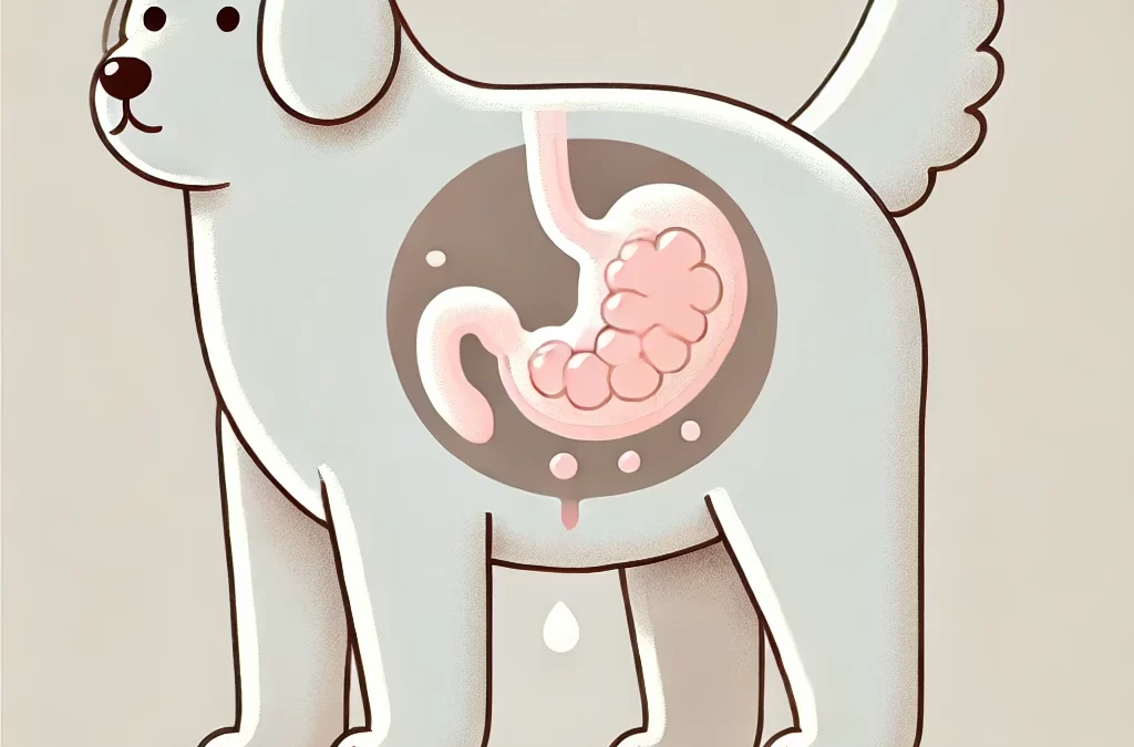
犬の腹部に大きな腫瘍が見つかった場合、飼い主は誰でも不安に思うでしょう。心配するのは当然ですが、考えられる原因、症状、治療の選択肢を理解することで、犬の健康について十分な情報に基づいた判断を下すことができます。
腹部腫瘤とは何ですか?
腹部腫瘤は、肝臓、脾臓、腸などの臓器、または周囲の組織から発生する可能性のある、胃の領域における異常な増殖です。これらの腫瘤は大きさがかなり異なり、良性(非癌性)または悪性(癌性)の場合があります。
腹部腫瘤の一般的な原因
- 良性腫瘍
- 脂肪腫: 一般的に無害な脂肪腫瘍。
- 嚢胞: 時間の経過とともに大きくなる可能性があるが、通常は危険ではない、液体で満たされた袋。
- 悪性腫瘍
- 血管肉腫:脾臓に発生する一般的な癌。
- リンパ腫:リンパ節に影響を及ぼし、腹部に転移する可能性があります。
- 癌腫:内臓の上皮組織から発生する腫瘍。
- その他の原因
- 膿瘍: 膿がたまったポケットにつながる感染症。
- 臓器肥大: 肝臓病などの病気は、臓器の肥大に似た症状を引き起こすことがあります。
注意すべき症状
早期発見は結果に大きな影響を与える可能性があります。次の点に注意してください。
- 腹部の腫れまたは膨張
- 体重減少または食欲減退
- 嘔吐または下痢
- 無気力または不快感の兆候
- 腹部の圧迫による呼吸困難
診断
腹部腫瘤の診断には獣医師の専門知識が必要です。手順には以下が含まれます。
- 身体検査: 腹部を触診して異常な腫瘍を検出する
- 画像検査: 超音波またはX線検査により腫瘍の大きさ、位置、性質を判定する
- 生検または穿刺吸引: 分析のための組織サンプルの収集
- Blood Tests: 関連する健康状態の確認
治療の選択肢
治療法は腫瘍の種類と場所によって異なります。
- 外科的除去: 手術可能な集団にとっての第一線となることが多い
- 化学療法または放射線療法: 悪性腫瘍、特に手術が不可能な場合に使用される
- 緩和ケア: 症状が治療できない場合は、快適さを維持することに重点を置きます
ペットの飼い主のための積極的な対策
- 定期検診: 定期的な獣医の診察は問題を早期に発見するのに役立ちます
- 症状を監視する: 犬の行動や食欲の変化に注意してください
- 適切な栄養: バランスの取れた食事は全体的な健康と回復をサポートします
- セカンドオピニオン: 複雑なケースについては、ためらわずに他の獣医に相談してください
犬の腹部の大きな腫瘤は深刻な問題ですが、適切なタイミングで獣医の診察と適切な管理を受ければ、多くの犬は良い結果を得ることができます。常に獣医と緊密に協力し、愛犬にとって最善の治療法を決定してください。
