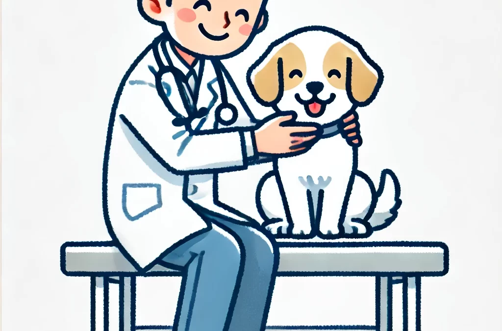
par TCMVET | Nov 29, 2024 | Cancer et tumeurs du chien
When faced with the emotional and financial burden of a dog’s tumor diagnosis, many pet owners feel overwhelmed by the costs associated with treatment. However, low-cost dog tumor removal is not only possible but also accessible with the right resources and a bit of creativity. This guide explores unique and practical ways to ensure your furry friend gets the care they need without straining your wallet.
Understanding Tumor Types and Necessity for Removal
Not all tumors require immediate surgical intervention. Some, such as lipomas (benign fatty tumors), may pose little to no threat to your dog’s health. Consulting with your veterinarian to assess the nature of the tumor is the first step toward making an informed decision. If removal is deemed necessary, explore cost-effective options to address the issue.
Creative Approaches to Affordable Dog Tumor Removal
Vet Schools: Learning and Saving
Veterinary teaching hospitals often provide lower-cost services as students, under the guidance of experienced professionals, perform procedures. While costs are reduced, the quality of care remains high, making this an excellent option for budget-conscious pet owners.
Non-Profit Veterinary Clinics
Many non-profits and animal welfare organizations offer subsidized veterinary care, including tumor removal. Research organizations in your area that support low-income pet owners, such as the Humane Society or SPCA branches, which may provide financial aid.
Crowdfunding Platforms
Platforms like GoFundMe or Waggle allow pet owners to share their dog’s story and raise funds for medical procedures. A heartfelt narrative and photos can encourage donors to contribute to your pet’s well-being.
Payment Plans with Local Vets
Some veterinary clinics are open to setting up payment plans for costly surgeries. This option lets you spread the cost over several months, easing the immediate financial burden.
DIY Recovery and Aftercare
While the surgery itself should always be performed by a licensed veterinarian, you can save money by managing post-operative care at home. Learn how to clean wounds, administer medications, and create a comfortable recovery space for your dog to minimize additional clinic visits.
Exploring Alternatives: When Surgery Isn’t an Option
If surgery is beyond your financial means or not recommended due to your dog’s health, alternative therapies can help manage the tumor. Some holistic options include:
- Herbal Supplements: Natural remedies like turmeric, frankincense, and TCM formulas such as Chuanxiong have been noted for their anti-inflammatory properties.
- CBD Oil: Full-spectrum CBD oil may help reduce inflammation and support overall well-being.
- Dietary Adjustments: Anti-cancer diets focusing on whole, low-carb, high-protein foods may slow tumor growth.
Preventative Measures to Avoid Future Costs
Preventative care can reduce the likelihood of tumors forming in the first place. Regular vet check-ups, a balanced diet, and avoiding exposure to carcinogens like tobacco smoke can promote a healthier life for your dog. Early detection is key; small tumors are generally easier and less expensive to treat than advanced ones.
Compassion and Ingenuity: The Keys to Affordable Care
Finding low-cost options for dog tumor removal requires resourcefulness and determination. By leveraging non-profits, community support, and alternative therapies, you can ensure your dog gets the care they need without financial hardship. Remember, your creativity and love for your pet can pave the way for compassionate and effective solutions.
Whether it’s through connecting with a network of affordable care providers or embracing holistic approaches, your dog’s health doesn’t have to come at an insurmountable cost. Let this journey remind us of the unwavering bond between pets and their owners—a relationship worth every effort.

par TCMVET | 28 novembre 2024 | Cancer et tumeurs du chien
Lorsque votre chien commence à boiter ou à se lécher les pattes de manière excessive, le coupable peut être un kyste interdigital, une affection à la fois courante et préoccupante. Bien que ces kystes soient généralement bénins, leur apparition peut parfois amener les propriétaires d'animaux à se demander s'il pourrait s'agir du signe d'un problème plus grave, comme un cancer. Examinons ce sujet avec un œil neuf, en explorant les distinctions, les liens potentiels et la meilleure façon d'aborder ces problèmes pour le bien-être de votre chien.
Que sont les kystes interdigitaux ?
Les kystes interdigitaux, également appelés furoncles, sont des bosses remplies de liquide qui se forment entre les orteils d'un chien. Ils résultent d'une inflammation des follicules pileux dans les espaces interdigitaux, souvent causée par :
- Trauma:Coupures ou égratignures sur les pattes.
- Allergies:Allergies environnementales ou alimentaires entraînant un léchage excessif.
- Prédispositions génétiques:Certaines races, comme les bouledogues et les labradors retrievers, sont plus sujettes à ce problème.
- Corps étrangers:Échardes ou débris incrustés dans la peau.
Bien que les kystes interdigitaux ne soient généralement pas cancéreux, leur nature récurrente peut provoquer une gêne, une infection et même une boiterie.
Les kystes interdigitaux peuvent-ils être liés au cancer ?
La réponse courte : rarement, mais pas totalement impossible.
La plupart des kystes interdigitaux sont bénins et non liés au cancer. Cependant, l'inflammation chronique causée par des kystes persistants peut potentiellement créer un environnement propice à des conditions plus graves au fil du temps. Ce phénomène, connu sous le nom de carcinogenèse induite par l'inflammation chronique, souligne comment une irritation prolongée peut augmenter le risque de modifications malignes dans les tissus.
De plus, dans de très rares cas, des bosses initialement identifiées comme des kystes peuvent en fait être quelque chose de plus grave, comme :
- Squamous Cell Carcinoma (SCC):Un type de cancer de la peau qui peut se développer dans les coussinets des pattes ou entre les orteils.
- Tumeurs à mastocytes (TMC):Ces tumeurs, bien que communément trouvées ailleurs, peuvent parfois apparaître dans des endroits inhabituels comme les pattes.
- Mélanome:Les mélanomes malins peuvent également se manifester près des coussinets des pattes et ressembler à des excroissances ressemblant à des kystes.
Comment différencier les kystes du cancer
Un diagnostic précis est essentiel. Voici ce à quoi il faut prêter attention :
- Forme et texture:Les kystes sont généralement mous, ronds et remplis de liquide. Les tumeurs peuvent être dures et irrégulières.
- Taux de croissance:Les kystes bénins se développent lentement, tandis que les tumeurs malignes se développent souvent rapidement.
- Couleur et ulcération:Les tumeurs cancéreuses peuvent être décolorées, ulcérées ou saigner spontanément.
- Réponse au traitement:Les kystes répondent souvent aux antibiotiques, aux médicaments anti-inflammatoires ou au drainage, alors que les tumeurs cancéreuses ne le font pas.
Les vétérinaires peuvent recommander des tests de diagnostic comme l’aspiration à l’aiguille fine (FNA) ou la biopsie pour confirmer si une croissance est bénigne ou maligne.
Soins holistiques et préventifs pour la santé des pattes
Même si un kyste n'est pas cancéreux, la prévention et les soins holistiques peuvent améliorer la qualité de vie de votre chien :
- Hygiène des pattes:Un nettoyage régulier réduit le risque d'intrusion d'objets étrangers dans les pattes de votre chien.
- Adaptations diététiques:Les acides gras oméga-3 et les suppléments anti-inflammatoires peuvent réduire l’inflammation et favoriser la santé de la peau.
- Natural Remedies: Products like TCMVET Baituxiao ou les crèmes à base de curcuma peuvent aider à réduire l’inflammation des kystes récurrents.
- Exercice modéré:Pour les chiens actifs sujets aux traumatismes, envisagez des terrains plus doux pour les promenades afin d'éviter les coupures et les écorchures.
Lorsqu'une intervention chirurgicale ou un traitement avancé est nécessaire
En cas de kystes persistants ou compliqués, une intervention chirurgicale peut être nécessaire pour retirer le tissu affecté. Dans de rares cas de suspicion de malignité, l'amputation de l'orteil affecté peut être nécessaire pour empêcher la propagation du cancer. Des thérapies avancées comme la chirurgie au laser ou la cryothérapie peuvent également fournir des solutions non invasives dans certains cas.
Le point à retenir : la connaissance est un pouvoir
Les kystes interdigitaux, bien que courants et généralement bénins, ne doivent jamais être ignorés. Rester vigilant, consulter votre vétérinaire et adopter une approche proactive peuvent faire toute la différence pour assurer la santé des pattes de votre chien. Et n'oubliez pas que même si le spectre du cancer se présente, une détection précoce et des traitements modernes offrent des résultats prometteurs.
Nos amis à fourrure comptent sur nous pour les soins et la protection. En comprenant les nuances de certaines pathologies comme les kystes interdigitaux, nous pouvons leur offrir les meilleures chances de vivre une vie longue, saine et heureuse.

par TCMVET | 28 novembre 2024 | Cancer et tumeurs du chien
When a beloved pet is diagnosed with a tumor, the emotional weight can feel overwhelming. Surgery often becomes the focal point of hope, but is it always the best option? Let’s explore the transformative journey of pet tumor surgery, the alternatives reshaping the landscape, and how holistic care is redefining healing for our furry companions.
A Historical Perspective: Surgery as a Lifesaver
In the early days of veterinary medicine, surgery was the definitive response to tumors. With advancements in technology, techniques such as laser surgery and robotic assistance have become common, making procedures safer and more precise. For example, oncological surgeries for pets today can achieve margins as small as a millimeter, preserving healthy tissue while removing the tumor.
However, the surgical path isn’t without its risks. Factors like the pet’s age, the size and location of the tumor, and underlying health conditions all influence the success rate. Despite its effectiveness in removing tumors, surgery often addresses only the symptom—not the root cause.
The Alternatives: A Growing Spectrum of Options
While surgery remains a cornerstone, alternative approaches are gaining traction:
- Cryochirurgie: Using extreme cold to freeze and destroy tumor cells, this method is less invasive and ideal for superficial growths.
- Thérapies ciblées: Innovations in veterinary medicine, such as immunotherapy and molecularly targeted drugs, are allowing for non-surgical tumor management. These treatments aim to shrink tumors or slow their progression.
- Natural Therapies: Herbal remedies like TCMVET Baituxiao and hemp-based supplements are becoming popular for their ability to support the immune system and inhibit tumor growth with minimal side effects.
- Soins palliatifs: For pets with inoperable tumors, comfort becomes the priority. Pain management, dietary adjustments, and physiotherapy play crucial roles in maintaining quality of life.
Weighing the Decision: To Cut or Not to Cut?
Choosing surgery or an alternative depends on multiple factors:
- Type de tumeur: Benign tumors may not necessitate immediate surgery, while malignant growths often require prompt action.
- Qualité de vie: Is the procedure likely to improve the pet’s well-being, or could it introduce undue stress and pain?
- Owner’s Goals: Some owners prioritize longevity, while others focus on comfort and holistic care.
Consultation with a veterinary oncologist is vital to ensure a tailored treatment plan that considers the pet’s unique needs.
Holistic Healing: Beyond the Scalpel
Post-surgical care is just as important as the procedure itself. Increasingly, holistic methods are being integrated into recovery plans:
- Thérapie nutritionnelle: Diets rich in antioxidants, omega-3 fatty acids, and cancer-fighting compounds are pivotal.
- Acupuncture and Massage: These therapies can alleviate pain, boost circulation, and accelerate healing.
- Soutien affectif: Pets, much like humans, benefit from a stress-free environment during recovery. Spending quality time, engaging in gentle play, and maintaining routines can uplift their spirits.
The Future of Pet Tumor Surgery
The field of veterinary oncology is evolving rapidly. Innovations such as AI-guided diagnostics and 3D-printed surgical tools promise even more precise and effective interventions. Additionally, research into the genetic basis of tumors in pets is paving the way for preventative strategies.
As these advancements unfold, the narrative around pet tumor surgery is shifting—from fear to hope, from reactive to proactive care.
Une dernière réflexion
Whether opting for surgery or exploring alternatives, the ultimate goal is always the same: giving our pets the best life possible. With compassion, informed decision-making, and access to cutting-edge care, we can navigate this challenging chapter and emerge stronger—together.
When faced with a tumor diagnosis, remember: You are your pet’s advocate and biggest champion. Every decision you make comes from love, and that makes all the difference.
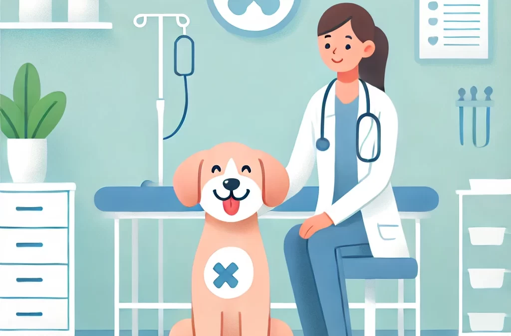
par TCMVET | Nov 27, 2024 | Cancer et tumeurs du chien
Tigilanol tiglate, a groundbreaking treatment for certain types of canine tumors, has been a game-changer in veterinary oncology. As pet owners explore this innovative therapy, many are concerned about its cost. But does the conversation about cost go beyond just the monetary figure? Let’s dive into a unique perspective on the value of tigilanol tiglate—looking not just at what it costs, but what it offers in return.
Understanding Tigilanol Tiglate
Developed from the seeds of the blushwood tree, tigilanol tiglate (marketed as Stelfonta®) offers a non-surgical solution to mast cell tumors in dogs. Administered via injection, it works by destroying tumor cells and encouraging wound healing, often with visible results within days. This innovative therapy is particularly appealing for dogs that are not candidates for surgery due to age, health, or tumor location.
The Financial Costs
The price of tigilanol tiglate can vary widely, depending on several factors:
- Tumor Size
The drug is priced based on dosage, which is determined by the tumor’s volume. Larger tumors require higher doses, increasing the cost.
- Veterinary Fees
The cost of administration includes pre-treatment assessments, sedation, the procedure itself, and follow-up care. These professional fees can vary by location and clinic.
- Soins post-traitement
While many dogs recover quickly, some may require additional wound management, which can add to the overall expense.
On average, the cost for tigilanol tiglate treatment ranges from $500 to $2,500 or more, depending on the above factors.
The Emotional Costs
While financial expenses are significant, the emotional toll of treating a beloved pet must also be considered. Tigilanol tiglate offers a compelling alternative to invasive surgery, reducing the stress and recovery time for both pets and their owners.
- Less Anxiety for Your Dog
Surgery often involves longer recovery periods and higher risks, particularly for older dogs. Tigilanol tiglate minimizes these challenges, offering a less invasive solution.
- Peace of Mind for Owners
Watching a tumor shrink in real-time can be an emotionally rewarding experience. For many, the cost is justified by the visible, immediate results.
Cost vs. Value
When evaluating the price of tigilanol tiglate, it’s essential to consider its value:
- Qualité de vie: The treatment focuses on preserving and enhancing the dog’s well-being without the risks associated with surgery.
- Time Saved: With a single injection, the treatment often eliminates the need for lengthy recovery periods.
- Emotional Relief: The ability to see rapid improvement can be priceless for pet owners struggling with the burden of their dog’s diagnosis.
Hidden Savings
While the upfront costs may seem high, tigilanol tiglate can offer indirect savings:
- Avoidance of surgery-related complications and their associated costs.
- Reduced need for ongoing treatments or medications for tumor management.
- Prevention of future tumor-related issues through early intervention.
Affording Tigilanol Tiglate: Tips for Pet Owners
For those concerned about affordability, here are some tips to explore:
- Pet Insurance: Check whether your policy covers advanced treatments like tigilanol tiglate.
- Payment Plans: Many veterinary clinics offer installment plans to help manage costs.
- Non-Profit Assistance: Organizations dedicated to pet care sometimes provide financial aid for critical treatments.
- Budgeting Ahead: Early financial planning for your pet’s healthcare can ease the burden of unexpected expenses.
Dernières pensées
The cost of tigilanol tiglate isn’t just a number—it’s a measure of hope, innovation, and care. While the treatment may not fit every budget, it offers unparalleled value for dogs and their families by providing a minimally invasive, effective option for managing mast cell tumors. For many, the question isn’t just “How much does it cost?” but rather, “What is the cost of not trying?”
As you navigate your dog’s treatment journey, remember that the price of care includes not only the dollars spent but also the moments you gain together. Tigilanol tiglate represents a future where more dogs can live healthier, happier lives—an investment in love, longevity, and companionship.
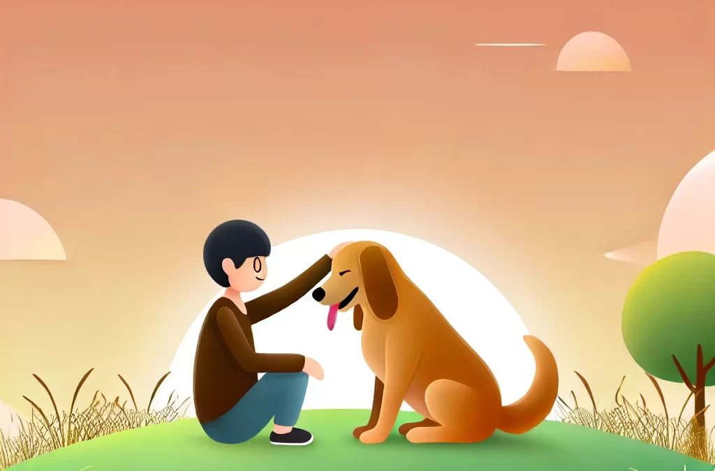
par TCMVET | Nov 27, 2024 | Cancer et tumeurs du chien
Deciding when to say goodbye to a beloved dog diagnosed with cancer is one of the hardest decisions any pet owner will face. While every case is unique, this article offers an alternative perspective on this emotionally charged topic—focusing on love, quality of life, and creating meaningful memories before letting go.
Understanding Your Dog’s Journey
Dogs with cancer, like humans, experience a range of physical and emotional states. Understanding their condition can help you make informed decisions:
- Pain Levels: Pain is often the first indicator. Despite advancements in palliative care, some dogs may experience persistent discomfort.
- Mobility Issues: Observe whether your dog can still enjoy activities like walking or playing. Loss of mobility may signify a decline in their quality of life.
- Appetite Changes: A sudden refusal to eat or drink could mean their body is shutting down.
- Emotional Changes: Dogs are emotional creatures. Notice if your dog seems withdrawn, anxious, or disinterested in their surroundings.
A New Framework: The “Five Joys” Approach
Instead of focusing solely on decline, consider this approach to assess your dog’s quality of life. Ask yourself:
- Eating: Does your dog still enjoy their favorite foods?
- Sleeping Comfortably: Are they resting without signs of pain or distress?
- Social Interaction: Do they seek companionship or enjoy being petted?
- Playing: Are they engaging in their favorite activities, even in a limited way?
- Exploring: Do they show interest in their environment?
When three or more of these joys are consistently missing, it may be time to consider euthanasia.
Saying Goodbye: A Holistic Approach
Saying goodbye doesn’t have to feel like the abrupt end of a chapter. Here are ways to honor your dog’s journey while easing the transition:
- Create a Bucket List
Celebrate your dog’s life by creating moments of joy. It could be as simple as a picnic in their favorite park or sharing a special treat.
- Focus on Comfort
Provide a calm, familiar environment. Use cozy bedding, gentle massages, and aromatherapy to ease their stress.
- Communicate with a Vet You Trust
A compassionate veterinarian can guide you in recognizing the signs of decline and help you plan a peaceful passing.
- Consider Home Euthanasia
Many pet owners opt for at-home euthanasia services, allowing their dog to pass in familiar surroundings, surrounded by loved ones.
- Preserve Their Legacy
Create a keepsake, such as a paw print mold or a scrapbook of cherished photos. This can help you process grief while celebrating your dog’s life.
Redefining the Final Goodbye
Euthanasia is not just a clinical decision—it’s an act of love. By choosing to let your dog go peacefully, you’re sparing them unnecessary suffering and honoring their dignity. Rather than focusing on “when to let go,” shift your perspective to “how to make their last moments meaningful.”
The Takeaway: It’s Okay to Grieve
Grief is a natural part of the process. It’s a testament to the deep bond you shared with your dog. Seek support from friends, family, or even online communities of pet lovers who understand what you’re going through.
In the end, the decision is deeply personal. Trust your instincts, honor your dog’s unique journey, and know that love, not time, defines your relationship with your pet.

par TCMVET | Nov 26, 2024 | Cancer et tumeurs du chien
When a pet parent hears the word “tumor,” it’s like a punch to the gut. The questions come flooding in: Is it cancerous? Will my dog be okay? And most importantly, what can I do to help? While modern veterinary medicine offers various treatments, many owners are turning to natural, holistic approaches to complement traditional care. Let’s explore some innovative and lesser-known options to help manage and potentially shrink tumors in dogs.
The Nature of Tumors in Dogs
Tumors in dogs can range from benign lipomas to malignant cancers like mast cell tumors or osteosarcomas. The treatment plan depends on the type, size, and stage of the tumor, but addressing it often involves a mix of conventional treatments (like surgery or chemotherapy) and supportive therapies to boost overall health.
But here’s the catch: not all treatments have to be invasive or synthetic. Nature has provided us with a treasure trove of resources that may help shrink tumors while supporting your dog’s well-being.
Nutritional Support: Food as Medicine
- Mushroom Powerhouses
Medicinal mushrooms like shiitake, reishiet turkey tail are rich in beta-glucans, which can help regulate the immune system and may slow tumor growth. Studies in veterinary oncology suggest that these fungi can reduce the progression of certain cancers. A sprinkle of powdered mushroom supplements in your dog’s food could be a game-changer.
- Golden Paste (Turmeric Blend)
Turmeric is known for its active compound, curcumin, a powerful anti-inflammatory and antioxidant. Curcumin has been shown to disrupt cancer cell growth in some studies. Mix turmeric powder with coconut oil and black pepper to create a dog-friendly golden paste.
- Acides gras oméga-3
Found in fish oil or flaxseed, omega-3s are natural anti-inflammatories that can slow the growth of tumors and support overall health. Add it to your dog’s meals for a simple yet impactful dietary boost.
Natural Supplements for Tumor Management
- Huile de CBD
Cannabidiol (CBD) has gained traction for its potential anti-tumor effects. It’s believed to induce apoptosis (programmed cell death) in cancer cells and reduce inflammation. Always choose a pet-specific CBD oil that’s free from THC and consult your vet for the right dosage.
- Thé Essiac
A blend of herbs including burdock root, slippery elm, and sheep sorrel, Essiac tea has long been used as a natural remedy for tumors. It’s available in liquid or capsule form and is believed to help detoxify the body and shrink abnormal growths.
- Chuanxiong (Szechuan Lovage)
A lesser-known but potent traditional Chinese herb, Chuanxiong has properties that improve circulation and reduce inflammation. Some holistic vets recommend it as part of an herbal protocol for managing tumors.
Thérapies holistiques
- Acupuncture
While not directly shrinking tumors, acupuncture can improve blood flow, reduce pain, and enhance the efficacy of other treatments. It’s a great addition to a multi-faceted care plan.
- Hyperbaric Oxygen Therapy (HBOT)
Tumors thrive in low-oxygen environments. Hyperbaric oxygen therapy saturates the body with oxygen, potentially slowing tumor growth and aiding in healing.
Ajustements du style de vie
- Dietary Overhaul
A low-carb, high-protein diet can starve certain tumors that rely on sugar for growth. Consider a raw or cooked diet tailored to your dog’s specific needs.
- Réduction du stress
Chronic stress can suppress the immune system, making it harder for your dog to fight off diseases. Ensure your dog’s environment is calm and enriched with activities they enjoy.
- Detoxifying the Environment
Reduce exposure to harmful chemicals like pesticides, synthetic air fresheners, and processed foods. These environmental toxins can burden your dog’s system, making recovery more challenging.
The Power of Combining Modern and Natural Approaches
While natural therapies offer incredible benefits, they’re not a standalone solution for all dogs. Partnering with a vet who understands integrative care is crucial. This ensures your dog receives the best of both worlds: the life-saving power of modern medicine and the gentle support of natural remedies.
A Hopeful Outlook
The journey of helping your dog through a tumor diagnosis can feel daunting, but remember: you’re not alone. With a blend of dietary changes, natural supplements, and holistic therapies, you can give your dog a fighting chance while improving their quality of life.
Sometimes, the smallest changes—a dash of turmeric, a spoonful of mushroom powder, or a drop of CBD—can make the biggest difference.