
par TCMVET | 21 janvier 2025 | Cancer et tumeurs du chien
Regarder un chien bien-aimé se battre contre un cancer en phase terminale est un voyage émotionnel et déchirant. Même si la guérison n’est peut-être pas possible, il existe des moyens d’améliorer son confort, de soulager la douleur et d’améliorer sa qualité de vie restante. Cet article explore des approches pratiques, compatissantes et même alternatives pour soutenir les chiens atteints d’un cancer en phase terminale.
1. Donner la priorité à la gestion de la douleur
Le soulagement de la douleur est la pierre angulaire des soins contre le cancer à un stade avancé. Les options conventionnelles de prise en charge de la douleur comprennent :
- Médicaments sur ordonnance contre la douleur – Les AINS (anti-inflammatoires non stéroïdiens) et les opioïdes (comme le tramadol) aident à gérer efficacement la douleur.
- Thérapies complémentaires – La gabapentine pour les douleurs nerveuses et l’amantadine comme antagoniste NMDA peuvent améliorer le confort lorsqu’elles sont associées à d’autres analgésiques.
- Huile de CBD et remèdes à base de plantes – De nombreux propriétaires d’animaux de compagnie se tournent vers l’huile de CBD à spectre complet, le curcuma et le boswellia pour aider à réduire l’inflammation et la douleur de manière naturelle.
2. Soutien nutritionnel pour renforcer la vitalité
Un régime alimentaire adapté aux personnes atteintes de cancer peut ralentir la progression de la maladie et améliorer le bien-être général. Tenez compte des éléments suivants :
- Protéine de haute qualité – Les viandes maigres et les poissons fournissent des acides aminés essentiels au maintien des muscles.
- Graisses saines – Les acides gras oméga-3 contenus dans l’huile de poisson aident à réduire l’inflammation et peuvent ralentir la croissance tumorale.
- Régime pauvre en glucides et riche en fibres – Les cellules cancéreuses se nourrissent de sucre, donc réduire l’apport en glucides peut aider à ralentir la progression.
- Suppléments naturels – Les champignons médicinaux comme la queue de dinde et le reishi, ainsi que les herbes chinoises comme l’astragale, peuvent soutenir le système immunitaire.
3. Thérapies holistiques pour le confort et la mobilité
Au-delà des médicaments et du régime alimentaire, les traitements holistiques peuvent apporter un soulagement supplémentaire :
- Acupuncture – Aide à réduire la douleur, à améliorer la mobilité et à stimuler l’équilibre énergétique.
- Massothérapie – Un massage doux améliore la circulation et soulage les raideurs.
- Hydrothérapie – La thérapie par l’eau chaude soulage la pression articulaire et favorise l’exercice doux.
- Soins énergétiques et Reiki – Certains propriétaires d’animaux de compagnie explorent la guérison énergétique pour se détendre et soulager le stress.
4. Créer un environnement confortable
Adapter l'espace de vie de votre chien peut faire une énorme différence :
- Literie souple et supports orthopédiques – Les lits en mousse à mémoire de forme réduisent les escarres et les douleurs articulaires.
- Contrôle de la température – Les chiens âgés et malades ont du mal à réguler leur température. Il est donc essentiel de les garder au chaud en hiver et au frais en été.
- Minimiser le stress – Un environnement calme et tranquille avec des odeurs familières peut aider à soulager l’anxiété et la douleur.
5. Gestion des problèmes digestifs et d'hydratation
Le cancer peut entraîner des nausées, des diarrhées et une perte d'appétit. Traiter ces symptômes contribue à améliorer le bien-être :
- Appetite Stimulants – Des médicaments comme la mirtazapine peuvent encourager l’alimentation.
- Bouillons d'os faits maison – Doux pour l’estomac, riche en nutriments et hydratant.
- Probiotiques et enzymes digestives – Soutient la santé intestinale et facilite la digestion.
- Hydratation fréquente – Assurer un apport adéquat en eau prévient la déshydratation et soutient la fonction rénale.
6. Soutien émotionnel et moments de complicité
Votre présence est l'un des plus grands réconforts pour un chien en phase terminale. Passez du temps ensemble :
- Participez à vos activités préférées – Une petite promenade, un trajet en voiture ou une séance de câlins tranquille peuvent apporter de la joie.
- Parlez à votre chien – Le son de ta voix est rassurant et apaisant.
- Soyez présent sans peur – Les chiens ressentent les émotions, donc maintenir une énergie paisible et aimante les aide à se sentir en sécurité.
7. Savoir quand dire au revoir
L’une des décisions les plus difficiles est de déterminer quand l’euthanasie est le choix le plus humanitaire. Considérez :
- L'échelle HHHHHMM – Développée par le Dr Alice Villalobos, cette évaluation de la qualité de vie mesure la douleur, la faim, l’hydratation, l’hygiène, le bonheur, la mobilité et plus de bons jours que de mauvais.
- Soins palliatifs vétérinaires et euthanasie à domicile – De nombreux vétérinaires proposent des services à domicile pour permettre une transition en douceur dans un environnement familier.
- Écouter votre chien – Des changements de comportement, une douleur persistante malgré les médicaments ou une perte totale d’appétit peuvent indiquer qu’il est temps.
Conclusion
Prendre soin d'un chien atteint d'un cancer en phase terminale est une expérience profonde remplie d'amour, de dévouement et de compassion. En mettant l'accent sur la gestion de la douleur, le soutien nutritionnel, les thérapies holistiques et le bien-être émotionnel, les propriétaires d'animaux de compagnie peuvent s'assurer que les jours restants de leur chien seront remplis de confort et de dignité. Que vous choisissiez les soins palliatifs ou que vous preniez finalement la difficile décision de dire au revoir, l'objectif reste le même : honorer le lien et leur offrir la meilleure qualité de vie possible.
par TCMVET | 20 janvier 2025 | Cancer et tumeurs du chien
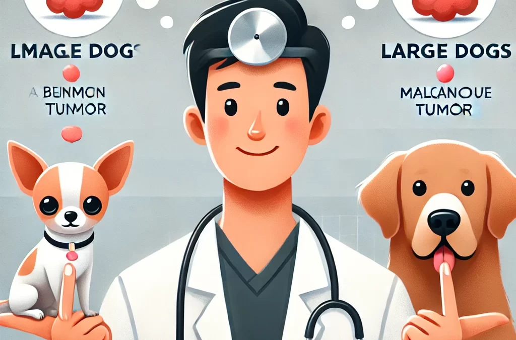
En ce qui concerne le développement du cancer et des tumeurs chez les chiens, la taille compte littéralement. Les petites et les grandes races ont des prédispositions génétiques, des taux métaboliques et des réponses immunitaires différents, qui contribuent tous aux variations dans la façon dont les tumeurs se développent, progressent et répondent au traitement. Si vous êtes un parent d'animal de compagnie préoccupé par la santé de votre chien, comprendre ces différences peut vous aider à prendre des décisions éclairées en matière de prévention, de détection précoce et de traitement.
1. Le facteur génétique : risques de tumeurs spécifiques à la race
Certains cancers sont plus fréquents chez certaines races, et la taille du chien joue souvent un rôle dans cette prédisposition.
- Grandes races:Les chiens comme les Golden Retrievers, les Dogues allemands et les Rottweilers sont plus susceptibles de développer ostéosarcome (cancer des os), hémangiosarcome (cancer des vaisseaux sanguins) et lymphomeCes cancers ont tendance à être agressifs et métastasent souvent rapidement.
- Petites races:Les races comme les caniches, les teckels et les chihuahuas sont plus sujettes à tumeurs bénignes telles que les lipomes et les papillomes, mais ils peuvent aussi se développer tumeurs mammaires et cancer de la vessie.
La différence ne réside pas seulement dans le type de tumeurs, mais également dans la manière dont ces cancers se comportent et répondent au traitement.
2. Taux de croissance et comportement de la tumeur
La progression tumorale varie considérablement entre les petits et les grands chiens en raison des différences de taux de croissance et de métabolisme cellulaire.
- Croissance plus rapide chez les grands chiens:Les races plus grandes grandissent rapidement en tant que chiots, et cette division cellulaire rapide peut contribuer à un risque plus élevé de développer tumeurs malignes plus tard dans la vie. Leurs tumeurs ont également tendance à être plus agressives.
- Croissance plus lente chez les petits chiens:Bien que les tumeurs chez les petites races puissent se développer plus lentement, elles restent préoccupantes. Tumeurs bénignes Les lipomes sont fréquents mais peuvent gêner la mobilité s'ils deviennent trop gros. De plus, les petits chiens peuvent encore développer des tumeurs malignes, telles que tumeurs à mastocytes, qui peut se propager si elle n’est pas traitée.
3. Espérance de vie et apparition de tumeurs
Les grands chiens ont tendance à avoir une durée de vie plus courte que les petits chiens, ce qui a un impact sur les délais de développement des tumeurs.
- Cancers à début précoce chez les grands chiens:Comme les grandes races vieillissent plus vite, elles sont plus susceptibles de développer un cancer à un plus jeune âge, souvent entre 6 à 8 ansCela signifie que les propriétaires doivent commencer les dépistages du cancer et les soins préventifs le plus tôt possible.
- Tumeurs à apparition tardive chez les petits chiens:Les petites races peuvent ne pas montrer de signes de cancer jusqu'à leur années seniors (10 ans et plus), ce qui signifie qu’une surveillance à long terme est essentielle.
La compréhension de ces délais peut aider les propriétaires d’animaux à planifier des examens vétérinaires au bon moment pour détecter précocement les tumeurs potentielles.
4. Défis en matière de diagnostic et de traitement
Lors du diagnostic et du traitement des tumeurs, la taille joue un rôle à la fois dans la détection et dans la capacité à effectuer des procédures.
- Considérations chirurgicales:Les grands chiens tolèrent mieux certaines interventions chirurgicales en raison de leur masse corporelle plus importante, mais l'ablation de tumeurs dans les os porteurs (comme dans le cas de l'ostéosarcome) peut être difficile. Les petits chiens, en revanche, peuvent avoir du mal à supporter les risques liés à l'anesthésie, surtout s'ils sont très petits.
- Différences entre chimiothérapie et médicaments:Le dosage de la chimiothérapie dépend du poids et les chiens de grande taille en ont souvent besoin. des doses de médicament plus élevées, ce qui augmente les coûts de traitement. Les petits chiens, bien que nécessitant des doses plus faibles, peuvent subir des effets secondaires plus forts en raison de leur système immunitaire fragile.
5. Stratégies de prévention et de détection précoce
Quelle que soit la taille du chien, une détection précoce est essentielle. Voici ce que les propriétaires de chiens peuvent faire :
- Examens vétérinaires de routine:Des examens réguliers permettent de détecter les tumeurs avant qu’elles ne deviennent trop grosses.
- Contrôles physiques à domicile: Passer vos mains sur le corps de votre chien chaque semaine peut aider à détecter des bosses inhabituelles.
- Dépistage du cancer selon la race:Les grandes races doivent subir des radiographies et des échographies précoces, tandis que les petites races peuvent bénéficier d'examens de la peau et de la vessie.
- Ajustements du régime alimentaire et du mode de vie:Une alimentation équilibrée, un exercice régulier et une gestion du poids peuvent contribuer à soutenir la santé globale et potentiellement à réduire les risques de cancer.
Dernières pensées
Bien que les tumeurs affectent aussi bien les petits que les grands chiens, leurs différences en termes de génétique, de comportement tumoral et d'options de traitement signifient que les propriétaires d'animaux ont besoin de stratégies de soins adaptées. Les grands chiens sont plus sujets aux cancers agressifs à un plus jeune âge, tandis que les petits chiens peuvent développer des tumeurs à croissance plus lente plus tard dans la vie. En comprenant ces distinctions et en donnant la priorité à la détection précoce, les propriétaires de chiens peuvent améliorer la qualité de vie de leurs animaux et potentiellement prolonger le temps qu'ils passent ensemble.
par TCMVET | 20 janvier 2025 | Cancer et tumeurs du chien
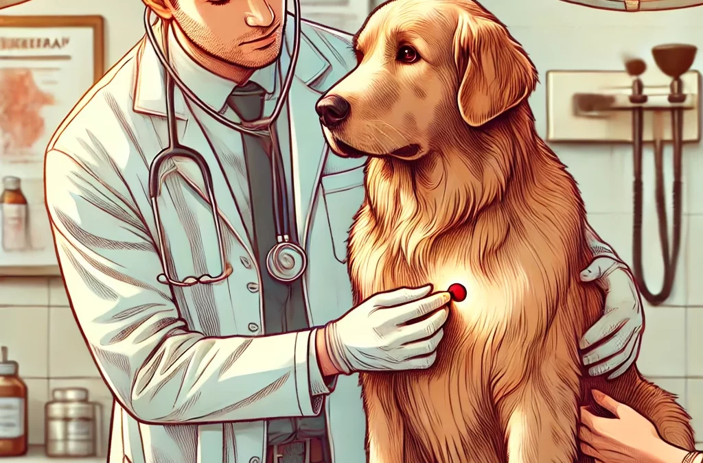
Imaginez la situation : vous venez de caresser le ventre de votre chien après une promenade bien remplie. Soudain, vos doigts effleurent une petite bosse que vous n'aviez jamais remarquée auparavant. Lancez la musique pleine de suspense : un envahisseur extraterrestre (lire : une tumeur maligne) a-t-il installé son campement, ou s'agit-il simplement d'un squatter inoffensif (une tumeur bénigne) qui se repose sous la peau de votre chien ? Avant de sonner l'alarme ou de l'ignorer, voyons comment décoder ces nouveaux locataires mystérieux dans le corps de votre ami à quatre pattes.
1. « Growth CSI » : des indices médico-légaux qui comptent
Imaginez-vous comme un détective dans une série policière à enjeux élevés. Votre travail consiste à rassembler toutes les preuves :
- Texture et mobilité:Les tumeurs malignes semblent souvent irrégulières et peuvent adhérer aux tissus environnants, tandis que les tumeurs bénignes ont tendance à être plus lisses et plus mobiles.
- Taux de croissance:Cette bosse gonfle-t-elle rapidement comme un ballon ou est-elle restée de la même taille au fil des semaines ou des mois ? Une croissance plus rapide peut indiquer une affection maligne.
- Symptômes associés:Des changements d’appétit, une léthargie, une perte de poids ou une douleur localisée peuvent être des signaux d’alarme supplémentaires qui nécessitent une attention immédiate.
Le point essentiel à retenir ? Ne tirez pas de conclusions hâtives en vous basant uniquement sur la sensation ressentie. Mais tenez à jour votre « carnet de détective » avec vos observations.
2. Renseignements scientifiques : les tests de diagnostic révèlent la vérité
Comme dans tout bon thriller, vous aurez besoin d'un partenaire qualifié pour résoudre l'affaire. Dans cet épisode, c'est votre vétérinaire de confiance, armé d'outils de haute technologie et d'un sens aigu du détail :
- Aspiration à l'aiguille fine (FNA):Un test rapide et peu invasif qui peut fournir des indices cellulaires immédiats. Considérez-le comme la collecte d'« empreintes digitales » de l'identité de la grosseur.
- Biopsie:Parfois, une analyse plus approfondie est nécessaire, comme l'analyse de l'ADN d'un suspect. Une biopsie permet d'examiner en profondeur la structure du tissu et le comportement cellulaire.
- Outils d'imagerie:Les rayons X, les ultrasons ou l'IRM agissent comme une loupe pour le détective, repérant des indices cachés sur les limites de la croissance et sa propagation potentielle.
3. Tumeur ou croissance bénigne ? Comprendre le verdict
Après avoir recueilli des preuves et effectué des tests, le verdict sera rendu. Les tumeurs malignes – nos « envahisseurs extraterrestres » – comportent un risque d’infiltration et de métastase. Cela nécessite souvent un traitement rapide, parfois agressif, qui peut inclure une intervention chirurgicale, une chimiothérapie ou une radiothérapie. Les tumeurs bénignes – nos « squatters inoffensifs » – grandissent généralement lentement et restent confinées, mais cela ne signifie pas que vous pouvez toujours les ignorer. Certaines tumeurs bénignes peuvent toujours appuyer sur des organes vitaux ou s’ulcérer au fil du temps, ce qui nécessite leur ablation ou une surveillance périodique.
4. Élaboration d’un plan directeur de traitement
Que le diagnostic soit celui d'un squatter inoffensif ou d'un envahisseur extraterrestre confirmé, avoir un plan d'action solide n'est pas négociable :
- Retrait chirurgical:Souvent la première ligne de défense, comme l’expulsion d’un mauvais locataire.
- Médicaments et thérapies:La chimiothérapie, la thérapie ciblée ou l’immunothérapie peuvent faire pencher la balance en faveur de votre chien si la tumeur est maligne.
- Ajustements du style de vie:Des régimes spécialisés aux routines d’exercices doux, un soutien holistique aide votre chien à se sentir au mieux tout au long du traitement.
- Surveillance continue:Considérez cela comme votre patrouille de périmètre pour vous assurer qu'aucun grumeau suspect ne revienne ou n'apparaisse ailleurs.
5. Célébrer les victoires et partager le parcours
La découverte d'une nouvelle bosse peut être un tournant effrayant dans l'histoire de votre animal de compagnie bien-aimé. Mais n'oubliez pas : toutes les bosses ne sont pas forcément un scénario pessimiste. Armé de vigilance et d'une approche proactive, vous pouvez transformer ce mystère « extraterrestre contre squatteur » en une intrigue secondaire gérable dans l'aventure de la vie de votre chien. Partagez les mises à jour avec votre vétérinaire, célébrez les petites victoires (une bosse stable, une opération réussie, un bon niveau d'énergie) et chérissez chaque mouvement de queue en cours de route.
Parce qu'en fin de compte, chaque histoire de détective qui mérite d'être racontée se termine par de l'espoir – et peut-être quelques friandises supplémentaires pour le meilleur acolyte du monde, votre chien.

par TCMVET | 18 janvier 2025 | Cancer et tumeurs du chien
Le cancer chez le chien est un défi de taille, souvent diagnostiqué trop tard pour une intervention efficace. Les outils de diagnostic traditionnels tels que les biopsies et l’imagerie ont leurs limites : ils peuvent être invasifs, coûteux ou incapables de détecter les tumeurs à un stade précoce. C’est là qu’entrent en jeu les biomarqueurs tumoraux : des signatures moléculaires trouvées dans le sang, l’urine ou les tissus qui offrent une approche révolutionnaire de l’oncologie canine. Les progrès de la médecine vétérinaire étant parallèles aux percées en oncologie humaine, la course est lancée pour développer des biomarqueurs fiables et non invasifs pour une détection précoce, une surveillance en temps réel et des stratégies de traitement personnalisées.
1. Que sont les biomarqueurs tumoraux et pourquoi sont-ils importants ?
Les biomarqueurs tumoraux sont des substances biologiques mesurables qui indiquent la présence, la progression ou la réponse au traitement du cancer. Il peut s'agir de :
- Protéines et enzymes:Des niveaux élevés de protéines spécifiques, telles que la protéine C-réactive (CRP) ou la thymidine kinase 1 (TK1), peuvent indiquer des tumeurs malignes.
- ADN tumoral circulant (ADNct):Des fragments d’ADN dérivés de tumeurs trouvés dans la circulation sanguine offrent des informations sur les mutations génétiques et la charge tumorale.
- Exosomes et microARN (miARN):De minuscules vésicules extracellulaires et des ARN non codants apparaissent comme des outils prometteurs pour la détection et le pronostic du cancer.
La capacité de détecter le cancer avant qu’il ne devienne cliniquement évident pourrait améliorer considérablement les résultats du traitement et la qualité de vie des chiens.
2. La révolution des biomarqueurs : du concept à l’application clinique
2.1. Détection précoce : la solution ultime pour changer la donne
Les cancers à un stade précoce sont souvent asymptomatiques, ce qui rend le dépistage systématique difficile. Les biomarqueurs peuvent combler cette lacune en identifiant les tumeurs malignes bien avant l'apparition des symptômes.
- CRP et TK1 spécifiques aux chiens:Des niveaux élevés ont été associés au lymphome, à l’hémangiosarcome et aux tumeurs des mastocytes.
- MicroARN sériques:Certains profils de miRNA sont fortement corrélés à l’ostéosarcome et aux tumeurs mammaires, ouvrant la voie à des tests sanguins de routine pour détecter les cas à haut risque.
2.2. Informations pronostiques : Prédire les résultats avec précision
Toutes les tumeurs ne se comportent pas de la même manière. Les biomarqueurs aident les vétérinaires à différencier les cancers agressifs des néoplasmes à croissance lente, ce qui permet d'élaborer des stratégies de traitement sur mesure.
- Ki-67 et PCNA (marqueurs de prolifération):Des niveaux d’expression élevés suggèrent une croissance tumorale rapide et un pronostic plus sombre.
- LDH (lactate déshydrogénase):Des taux élevés de LDH indiquent souvent des métastases dans l'hémangiosarcome, guidant l'intensité du traitement.
2.3. Suivi thérapeutique : ajustements de traitement en temps réel
Les biomarqueurs permettent un suivi non invasif de la réponse tumorale, permettant aux vétérinaires d'ajuster les traitements de manière dynamique.
- ADN tumoral circulant (ADNct):La surveillance des niveaux d’ADNct peut indiquer dans quelle mesure un chien répond à la chimiothérapie ou à la radiothérapie.
- Profilage des exosomes:Les changements dans la composition de la cargaison exosomale après le traitement fournissent des indices sur la maladie résiduelle et le risque de rechute.
3. Des technologies de pointe façonnent l'avenir de l'oncologie canine
3.1. L'intelligence artificielle (IA) rencontre les biomarqueurs
Les outils de diagnostic basés sur l’IA sont désormais formés pour analyser les profils de biomarqueurs, offrant des évaluations quasi instantanées et extrêmement précises. Imaginez un test sanguin basé sur l’IA qui prédit le risque de cancer avant l’apparition des signes cliniques !
3.2. Biopsie liquide : la fin des diagnostics invasifs ?
La biopsie liquide, qui détecte l'ADNct et les marqueurs exosomal, est sur le point de révolutionner le diagnostic du cancer. Contrairement aux biopsies traditionnelles, elle offre un aperçu en temps réel et peu invasif de l'évolution de la tumeur.
3.3. Médecine personnalisée pour chiens
À mesure que la recherche sur les biomarqueurs progresse, les vétérinaires pourraient bientôt avoir accès à une prise de décision basée sur les biomarqueurs, sélectionnant la meilleure chimiothérapie, immunothérapie ou traitements ciblés en fonction du profil tumoral unique d'un chien.
4. Défis et considérations éthiques
Malgré leurs promesses, les diagnostics basés sur les biomarqueurs se heurtent à des obstacles :
- Problèmes de normalisation:Les niveaux de biomarqueurs peuvent varier en fonction de la race, de l’âge et des maladies concomitantes.
- Coût vs. Accessibilité:Les tests de biomarqueurs avancés sont encore coûteux et peu disponibles.
- Faux positifs et négatifs:Aucun test de biomarqueur n’est infaillible ; des améliorations supplémentaires sont nécessaires pour garantir sa fiabilité.
5. Conclusion : l’aube d’une nouvelle ère dans le traitement du cancer canin
Les biomarqueurs tumoraux ne sont plus seulement des outils théoriques : ils deviennent rapidement indispensables au diagnostic, au pronostic et au traitement du cancer canin. En adoptant cette révolution moléculaire, la médecine vétérinaire entre dans un avenir où le cancer est détecté plus tôt, traité avec plus de précision et surveillé avec une précision sans précédent.
À mesure que la technologie évolue, le rêve d’un simple test sanguin permettant de dépister plusieurs cancers canins pourrait bientôt devenir réalité, offrant aux chiens et à leurs propriétaires le précieux cadeau de plus de temps et d’une meilleure qualité de vie.

par TCMVET | 18 janvier 2025 | Cancer et tumeurs du chien
Le monde de l’oncologie vétérinaire connaît un changement radical alors que les technologies de pointe et les recherches visionnaires convergent pour transformer notre approche des tumeurs cérébrales canines. Alors que les méthodes de diagnostic conventionnelles et les modalités de traitement traditionnelles guident depuis longtemps les praticiens, une nouvelle ère d’innovation promet de redéfinir les résultats cliniques et d’améliorer notre compréhension de ces pathologies complexes. Ci-dessous, nous explorons comment les outils de diagnostic de pointe, l’intelligence artificielle et l’influence croissante de la radiochirurgie stéréotaxique (SRS) repoussent les limites de la neuro-oncologie canine.
- Des symptômes à la suspicion : l’évolution du paysage diagnostique
1.1. Reconnaître l'inhabituel
Par le passé, la détection des tumeurs cérébrales canines reposait sur la reconnaissance de signes neurologiques subtils, tels qu’une inclinaison persistante de la tête, une ataxie et des changements de comportement ou d’appétit. Bien que ces signaux d’alarme restent cruciaux, l’imagerie avancée et l’analyse des données offrent une perspective plus nuancée. Les praticiens sont désormais mieux à même de faire la différence entre les états inflammatoires, les infections et les néoplasmes grâce à l’imagerie haute résolution et à des algorithmes de diagnostic perfectionnés.
1.2. L'essor de l'imagerie avancée
• IRM à haut champ : considérée comme la référence absolue pour la visualisation des lésions intracrâniennes, l'IRM à haut champ fournit des images détaillées des tissus mous et des limites des lésions. Les dernières séquences d'IRM, notamment l'IRM fonctionnelle (IRMf) et l'imagerie du tenseur de diffusion (ITD), peuvent approfondir la biologie tumorale, révélant des changements microstructurels avant l'apparition d'anomalies macroscopiques.
• Spectroscopie par résonance magnétique (SRM) : la SRM offre des informations au niveau moléculaire en évaluant les changements métaboliques au sein de la tumeur. Des pics de choline et de lactate élevés, par exemple, peuvent servir de signes avant-coureurs d'une tumeur maligne ou d'une croissance agressive.
• Analyse d'images assistée par IA : des algorithmes innovants basés sur l'intelligence artificielle permettent de détecter et de quantifier la croissance tumorale avec une rapidité et une précision remarquables. Ces outils peuvent intégrer des données cliniques, des marqueurs d'imagerie et des résultats histopathologiques pour prédire la progression probable d'une tumeur ou sa réponse au traitement.
1.3. Biopsie et au-delà
Bien que la technologie d’imagerie ait considérablement évolué, la confirmation histopathologique reste un pilier du diagnostic définitif. Les techniques de biopsie stéréotaxique minimisent le caractère invasif, réduisant ainsi les complications et accélérant la guérison. Dans un avenir proche, la biopsie liquide (analyse des cellules tumorales circulantes ou de l’ADN tumoral dans la circulation sanguine) pourrait réduire encore davantage le recours aux procédures invasives, ouvrant la voie à une surveillance tumorale en temps réel et à des ajustements dynamiques du traitement.
- Le saut quantique : la radiochirurgie stéréotaxique
2.1. Briser le moule de la radiothérapie conventionnelle
Pendant des décennies, la radiothérapie externe a été l'approche de facto pour traiter les tumeurs cérébrales inopérables ou difficiles à traiter chirurgicalement chez les chiens. Bien qu'efficace dans certains cas, elle impliquait souvent plusieurs séances sur plusieurs semaines. C'est là qu'intervient la radiochirurgie stéréotaxique (SRS), une technique de précision qui délivre une dose de rayonnement concentrée à la tumeur en une ou quelques séances seulement, minimisant ainsi les dommages aux tissus sains environnants.
2.2. Caractéristiques du SRS
• Précision extrême : l’imagerie avancée et la planification informatisée du traitement garantissent que le faisceau de rayonnement cible uniquement la tumeur, épargnant les structures environnantes.
• Séances de traitement réduites : De nombreux protocoles SRS canins nécessitent moins de visites, réduisant ainsi le stress pour l’animal et son propriétaire.
• Soulagement rapide des symptômes : la radiothérapie à haute dose réduit souvent la tumeur plus rapidement, offrant un contrôle plus rapide des symptômes par rapport à la radiothérapie fractionnée traditionnelle.
• Effets secondaires minimes : l’approche ciblée se traduit par moins de complications liées aux radiations, telles que l’irritation cutanée ou la perte de cheveux.
2.3. Équipement de pointe
Les hôpitaux vétérinaires utilisent de plus en plus de systèmes autrefois réservés à la médecine humaine, comme les unités Gamma Knife et CyberKnife. Ces appareils s'appuient sur des centaines de faisceaux de rayonnement convergents ou sur un bras robotisé capable de délivrer des doses élevées de rayonnement sous plusieurs angles, garantissant ainsi une précision et un contrôle inégalés.
2.4. Intégration du SRS à d'autres modalités
La radiochirurgie stéréotaxique n’est plus une procédure isolée. De nombreux spécialistes préconisent une approche multimodale, associant :
• Chimiothérapie ou thérapie ciblée pour lutter contre les maladies microscopiques et les métastases à distance.
• Immunothérapie pour améliorer la capacité innée du corps à détecter et à détruire les cellules cancéreuses.
• Soutien nutritionnel et réadaptation pour améliorer le bien-être général, accélérer la récupération et maintenir la masse musculaire.
- La voie à suivre : défis et opportunités
3.1. Considérations financières et logistiques
Les technologies de pointe, comme les appareils de radiothérapie spécialisés, nécessitent des investissements financiers importants. Par conséquent, tous les centres vétérinaires ne peuvent pas proposer la radiothérapie sélective, ce qui limite l'accessibilité. Cependant, à mesure que la technologie évolue et que de plus en plus de cliniques adoptent des équipements de pointe, les coûts pourraient diminuer.
3.2 Repousser les limites de la recherche
Les données sur les résultats à long terme et les essais cliniques à grande échelle restent relativement rares en médecine vétérinaire. En cultivant des collaborations multidisciplinaires entre vétérinaires, oncologues, radiologues et physiciens médicaux, le domaine peut recueillir des preuves solides sur la sécurité, l'efficacité et les protocoles optimaux de la SRS chez les chiens.
3.3. Médecine personnalisée et au-delà
Le profilage moléculaire, les tests génétiques et les dossiers médicaux numériques fusionnent pour créer un environnement médical plus personnalisé dans les soins vétérinaires. Les innovations futures pourraient intégrer le suivi des biomarqueurs en temps réel pour adapter les stratégies de traitement à la volée, ce qui pourrait révolutionner la neuro-oncologie canine d'une manière que nous pouvons à peine imaginer aujourd'hui.
- Conclusion
Le diagnostic et le traitement des tumeurs cérébrales chez les chiens n’ont jamais été aussi sophistiqués, ni aussi prometteurs. La fusion de l’imagerie de haute précision, de l’analyse basée sur l’IA et de la radiochirurgie stéréotaxique redéfinit les possibilités en oncologie vétérinaire. Bien que des défis importants demeurent – de la garantie d’une accessibilité généralisée à la collecte de preuves à grande échelle –, l’élan collectif laisse entrevoir un avenir meilleur pour les patients canins atteints de tumeurs cérébrales.
En embrassant ces nouvelles frontières, nous élevons non seulement le niveau de soins en neuro-oncologie canine, mais aussi la mission plus large de la médecine vétérinaire elle-même : maximiser la santé, le confort et la longévité de nos chers compagnons. Alors que la SRS et d’autres thérapies révolutionnaires gagnent du terrain, ce qui semblait autrefois une possibilité lointaine devient rapidement la nouvelle norme, offrant aux chiens atteints de tumeurs cérébrales une seconde chance de vie vraiment remarquable.

par TCMVET | 17 janvier 2025 | Cancer et tumeurs du chien
Découvrir que votre chien est atteint d'une tumeur peut être une expérience pénible. L'une des premières questions que se posent les propriétaires d'animaux de compagnie est : « Depuis combien de temps mon chien est-il atteint ? » La réponse dépend de divers facteurs, notamment du type de tumeur, de son emplacement, de son stade, des options de traitement et de l'état de santé général du chien. Comprendre ces éléments peut vous aider à prendre des décisions éclairées et à fournir les meilleurs soins possibles à votre compagnon à quatre pattes.
Types de tumeurs et leur impact sur l'espérance de vie
Toutes les tumeurs ne mettent pas la vie de votre chien en danger. Certaines sont bénignes et n'affectent pas nécessairement sa durée de vie, tandis que d'autres sont malignes et peuvent se propager de manière agressive.
1. Tumeurs bénignes
Les tumeurs bénignes, comme les lipomes (tumeurs graisseuses) et les adénomes sébacés, n'ont généralement pas d'impact sur la durée de vie d'un chien, à moins qu'elles n'interfèrent avec les mouvements ou le fonctionnement des organes. Dans de nombreux cas, les chiens peuvent vivre une vie normale sans traitement, bien qu'une ablation chirurgicale puisse être envisagée pour plus de confort.
2. Tumeurs malignes (tumeurs cancéreuses)
Les tumeurs malignes présentent un risque plus grave car elles peuvent envahir les tissus environnants et métastaser vers d'autres organes. Les tumeurs malignes courantes chez le chien comprennent :
- Tumeurs à mastocytes (TMC) : La maladie peut se développer lentement ou être agressive. Avec une élimination précoce, de nombreux chiens continuent à vivre pendant des années, mais les cas avancés peuvent réduire considérablement leur durée de vie.
- Ostéosarcome (cancer des os) : Très agressif, il nécessite souvent une amputation et une chimiothérapie. Sans traitement, la durée de survie est généralement de 3 à 6 mois, mais avec un traitement, certains chiens vivent 1 à 2 ans ou plus.
- Lymphome : L'un des cancers canins les plus courants. Grâce à la chimiothérapie, la rémission peut prolonger la vie d'un chien de 1 à 2 ans, mais sans traitement, la durée de survie est généralement de 1 à 2 mois.
- Hémangiosarcome : Cancer à propagation rapide affectant la rate, le cœur ou le foie. Le pronostic est mauvais, la survie pouvant aller de quelques semaines à quelques mois, même avec un traitement.
- Tumeurs mammaires : Si la maladie est détectée à temps et retirée chirurgicalement, de nombreux chiens se rétablissent bien. Cependant, les cas malins qui se sont propagés réduisent considérablement l'espérance de vie.
Facteurs clés affectant la durée de survie d'un chien
Plusieurs variables influencent la durée de vie d’un chien atteint d’une tumeur :
1. Détection et diagnostic précoces
Plus tôt une tumeur est identifiée, meilleures sont les chances d'un traitement efficace. Des contrôles vétérinaires réguliers et une surveillance des grosseurs, de la perte de poids ou des changements de comportement peuvent conduire à une intervention précoce, ce qui peut prolonger considérablement l'espérance de vie.
2. Type, localisation et stade de la tumeur
La taille, l'emplacement et la propagation de la tumeur déterminent l'impact qu'elle aura sur la santé de votre chien. Les tumeurs affectant les organes vitaux ou le système nerveux ont tendance à être plus dangereuses, tandis que les tumeurs cutanées ou celles des membres peuvent être plus faciles à gérer.
3. Options de traitement et réponse
Le traitement affecte considérablement la durée de survie. Les options courantes comprennent :
- Chirurgie : Peut éliminer complètement les tumeurs bénignes et certains cancers localisés, conduisant potentiellement à une guérison complète.
- Chimiothérapie : Souvent utilisé pour traiter des cancers comme le lymphome. Même s'il ne guérit pas le cancer, il peut prolonger la vie d'un chien et améliorer sa qualité de vie.
- Radiothérapie : Aide à réduire les tumeurs qui ne peuvent pas être retirées chirurgicalement.
- Soins holistiques et palliatifs : Les remèdes naturels, les changements alimentaires et la gestion de la douleur peuvent améliorer le confort et le bien-être des chiens à un stade avancé.
4. Santé générale et âge
Un chien jeune avec un système immunitaire fort est plus susceptible de bien réagir au traitement qu'un chien plus âgé souffrant d'autres problèmes de santé sous-jacents. Maintenir un mode de vie sain avec une alimentation équilibrée et de l'exercice peut favoriser de meilleurs résultats.
5. Considérations relatives à la qualité de vie
Au-delà de la survie, la qualité de vie d'un chien est un facteur crucial. Surveiller le niveau de douleur, l'appétit, la mobilité et les niveaux d'énergie peut aider à déterminer la meilleure marche à suivre. Dans certains cas, l'euthanasie humaine peut être l'option la plus douce si un chien souffre.
Comment aider un chien atteint d'une tumeur
1. Travaillez en étroite collaboration avec un vétérinaire
Des contrôles réguliers, des tests de diagnostic et des plans de traitement personnalisés peuvent aider à maximiser la durée de vie et le confort de votre chien.
2. Proposer une alimentation riche en nutriments
Un régime alimentaire adapté au cancer, riche en protéines maigres, en acides gras oméga-3 et en antioxydants, peut soutenir le système immunitaire et la santé globale.
3. Gérer la douleur et l’inconfort
La gestion de la douleur, que ce soit par des médicaments, de l’acupuncture ou des suppléments à base de plantes, peut grandement améliorer le bien-être d’un chien.
4. Surveiller les changements de comportement
Remarquer des signes tels qu’une léthargie accrue, une perte d’appétit ou des difficultés respiratoires peut vous aider à ajuster vos stratégies de soins en temps opportun.
5. Envisager des thérapies alternatives
Certains propriétaires d'animaux de compagnie explorent la médecine traditionnelle chinoise, l'huile de CBD ou d'autres traitements holistiques pour soutenir la santé de leur chien parallèlement aux traitements conventionnels.
Dernières pensées
Un diagnostic de tumeur chez le chien ne signifie pas nécessairement que la fin est proche. De nombreux chiens vivent heureux pendant des mois, voire des années, grâce à des soins, un traitement et des ajustements de style de vie appropriés. La clé est la détection précoce, le traitement approprié et le maintien d'une qualité de vie élevée. Chaque chien est différent et en travaillant en étroite collaboration avec un vétérinaire, vous pouvez prendre les meilleures décisions pour garantir que le temps restant de votre animal soit aussi confortable et épanouissant que possible.
Souhaitez-vous en savoir plus sur des types de tumeurs spécifiques, des options de traitement ou des approches de soins holistiques ?
